Ankle Pain
Peroneal Tendon Tear
Read More >

Minute Read
|Posted 4 years ago
|Last updated: 17/06/2023
|by James McCormack

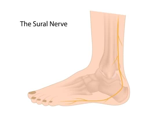
The Sural nerve is a primary sensory nerve which means it is the type of nerve that supplies sensation to the skin. The Sural Nerve can detect foot position, temperature, pain, vibration and touch as a sensory nerve.
The sural nerve is formed from the union of two smaller sensory nerves: the medial sural cutaneous nerve (a branch of the tibial nerve) and the lateral sural cutaneous nerve (a branch of the common fibular nerve).
The sural nerve courses distally along the posterolateral aspect of the leg between the heads of the gastrocnemius muscles in the posterior knee, parallel to the small saphenous vein. When the sural nerve reaches the ankle, it wraps around the lateral retro-malleolar region outside the peroneal tendons before splitting into two branches at the level of the 5th metatarsal.
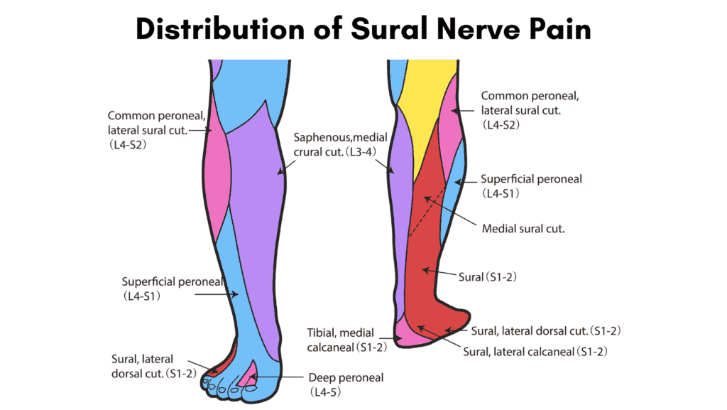
The sural nerve provides sensation to the lower 1/3 of the outer leg, outer heel and the outer aspect of the foot.
As the Sural Nerve is a superficial nerve, it can be easily irritated from muscular or fascial entrapment, ankle sprains, and being overstretched. When the sural nerve becomes inflamed, it is called Sural Neuritis, which often results in a burning pain in the lateral shin or the lateral foot (foot sural nerve pain).
Patients often describe their pain as constant pain that can be made worse with activity but remain present at rest. The constant pain at rest differentiates the symptoms of Sural Nerve pain from most other mechanical conditions, where pain normally eases with rest.
Sitting for long periods or driving can often exacerbate the symptoms of sural nerve pain as it places the nerve under tension.
Sural Nerve Pain is commonly misdiagnosed as the following conditions:
One of the most common causes of Sural Nerve Pain is a sprained ankle where the foot inverts at speed, stretching the Sural Nerve and causing it to become inflamed and painful.
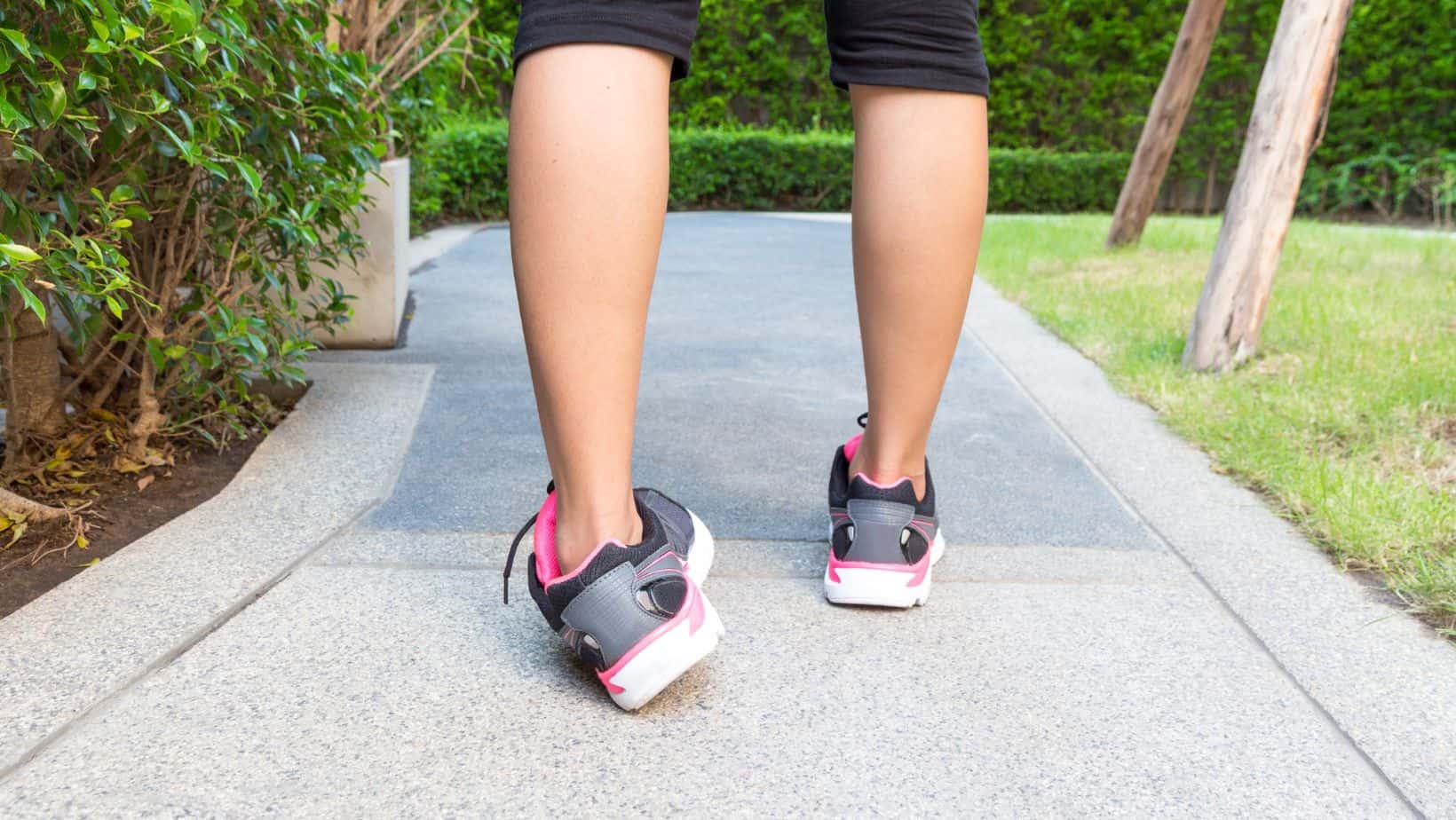
Tight shoes or ill-fitting ski boots can directly compress the Sural Nerve leading to a Sural Nerve Entrapment, or thickening of muscular tissue or fascia around the Sural nerve can irritate it.
Other causes of Sural Nerve Pain include irritation from metalwork after orthopaedic surgery or diabetes-related neuropathy, popliteal nerve entrapment, exertional compartment syndrome of the lower leg or a ganglia at the ankle.
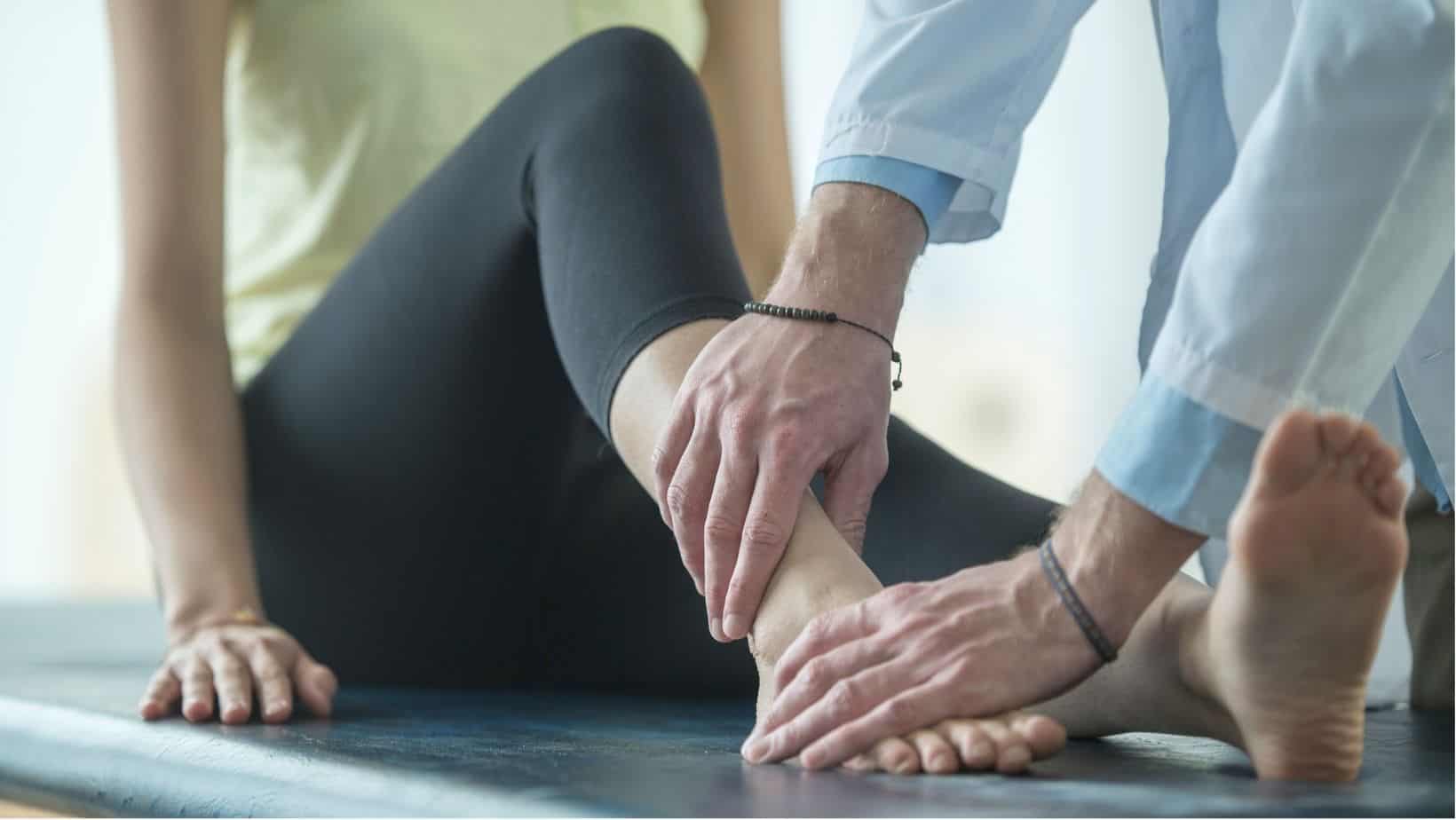
Sural Nerve Pain is usually diagnosed in a clinic by a Physical Therapist using a combination of clinical tests, such as a sural nerve tension test, a modified version of a straight leg raise test. Tapping (Tinel’s test) of the nerve can elicit the patient’s symptoms while it may be tender to palpate along the anatomical pathway of the Sural Nerve.
In most cases, it is challenging to identify Sural Nerve pain on a scan. There may be some signs of inflammation on an MRI scan, but in most cases, an MRI will return as ‘normal’.
On Ultrasound, Neuropathy will present as thickening and hypoechogenicity of the nerve. In contrast, a nerve tumour will present as a mass in continuity with the nerve or branch with increased doppler. Also, nerve transection from trauma will appear as an interruption of the nerve.
Treatment for Sural Nerve Pain is typical with Physical Therapy involving the ankle joint, foot and fibula mobilisation. Soft tissue release may be used on the surrounding tissues of the Sural nerve, such as the calf muscle, peroneal muscles and proximally on the lateral hamstring muscles. Advice on footwear and, in some cases, insoles can be helpful.
Exercises to relieve Sural Nerve pain include the Sural Nerve Glider and gentle stretching of the calf, hamstring and gluteal muscles.
To perform the sural nerve glider:
In most cases, manual therapy combined with the appropriate rehabilitation exercises is sufficient to relieve Sural Nerve Pain. In rare instances, a steroid injection may be required to reduce inflammation within the nerve and a period of 1-2 weeks in a walker boot may be recommended to allow the injection to take maximum effect.
This is not medical advice. We recommend a consultation with a medical professional such as James McCormack. He offers Online Physiotherapy Appointments weekly.