Orthotics and Insoles
Insoles For Flat Feet
Read More >

Minute Read
|Posted 4 years ago
|Last updated: 14/11/2023
|by James McCormack

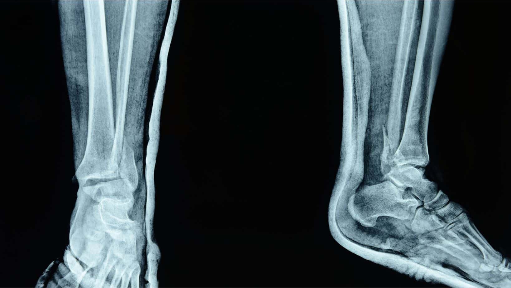
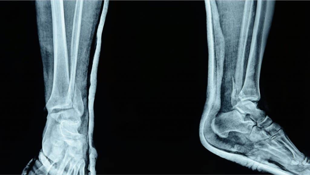
The ankle joint consists of 3 bones: the medial malleolus, lateral malleolus, and the talus bone. The lateral malleolus sits on the outside of the ankle joint and absorbs approximately 10% of your weight in standing.
The lateral malleolus is the most common of the ankle joints to fracture. While fractures are generally stable, traumatic events such as a fall or trip can lead to an unstable fracture.
This article will discuss the most common types of Lateral Malleolus Fractures, conservative treatment, and surgical treatment.
Related Article: Talus Fractures
In a study of 9767 patients with ankle fractures over 9 years, it was found that lateral malleolus fractures are the most common ankle fracture at a rate of 55%. There doesn’t appear to be much difference in the likelihood of developing a lateral malleolus fracture between genders; however, it appears more prevalent in females as they age.
This is a fracture at the ankle level of the lateral malleolus but may extend up the fibula. It can involve the ligaments attaching to the fibula and can be stable or unstable. Weber B fractures are generally stable fractures that can be managed conservatively with immobilisation and physical therapy.
This is when a gap appears in the bone at the site of the fracture and these often require surgical repair
This is a type of fracture involving the growth plate and is common in children
This is where the ligament is pulled off the bone and in doing so it can pull some flakes or fragments of the bone off with it.
The syndesmosis attached the tibia and fibula together, this becomes torn or compromised with this type of fracture.
A consultation with a Physical Therapist, Podiatrist, or Sports Medicine Doctor is recommended if you have any of the symptoms of a Lateral Malleolus Fracture. A clinical interview of your symptoms alongside a clinical examination can often be sufficient to achieve a diagnosis for your therapist. A clinical examination will often involve a hop test and tap test.
This is usually followed by imaging to rule out other conditions and to confirm the diagnosis. An X-ray is the primary imaging model, but if this returns, as usual, a referral for an MRI or CT scan may be required. A study of 123 patients with lateral and mortise x-ray views is 95% accurate in diagnosing ankle fractures as anteroposterior, lateral and mortise views.
A stable Lateral Malleolus Fracture is usually managed with 4-6 weeks in a walker boot. Historically, a cast was used, but the walker boot allows for greater function and equivalent healing response.
During this period, Physical Therapy can begin to maintain the mobility of the surrounding tissues, such as the Calf, Peroneal, and Posterior Tibialis muscles.
Upon boot removal, rehabilitation can last for 4-6 weeks. This involves strengthening and mobility exercises of the foot and ankle. Graded exposure to impact activities and return to play protocols if you wish to return to sport. Rehabilitation exercises are similar to that of a sprained ankle.
Taping and/or an ankle brace may be used when returning to activities that require a change of support, especially for those with unstable ankles. Surgery is not usually required for a stable fracture.
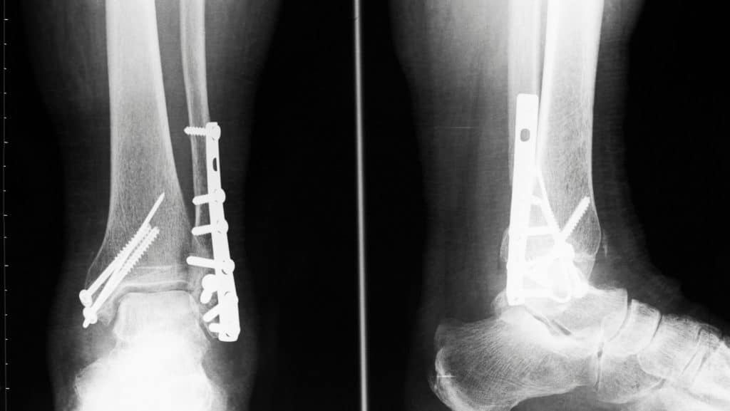
For unstable Lateral Malleolus Fractures or a malunion, surgery may be required. This is often an ORIF involving metalwork such as a plate stabilising the broken bone. You may require the use of a walker boot after surgery, and rehabilitation with a Physical Therapist can last for 6-12 weeks.
Post-operative complications include nerve damage if a nerve is cut during the procedure, but these surgeries are generally very successful overall.
You can still walk wearing an aircast boot with a Lateral Malleolus Fracture. Aircast boots are designed for walking short distances, and this should be indicated by your pain levels.
If your Lateral Malleolus Fracture is too painful to walk in a boot, try partial weight bearing using crutches instead.
This article is written by James McCormack, a Lower Limb Specialist who is an expert in treating Ankle Fractures.
This is not medical advice. We recommend a consultation with a medical professional such as James McCormack if you are experiencing any of the symptoms discussed in this article. James offers Online Physiotherapy Appointments weekly and face-to-face appointments in his London clinic.