
Knee Pain
Pes Anserine Bursitis
Read More >

Minute Read
|Posted 4 years ago
|Last updated: 04/12/2023
|by James McCormack

Meniscus tears are a common cause of knee pain. Tears are more common to the medial meniscus, on the inside of the knee, but can also occur on the lateral meniscus. It is common to have a meniscus injury in conjunction with a ligament sprain such as injury to the medial collateral ligament or anterior cruciate ligament.
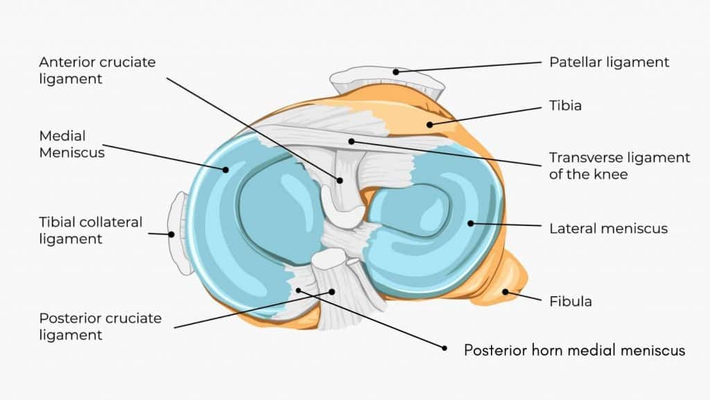
The meniscus is a thick cartilage structure that sits between the bones of the knee. It has the shape of two C’s, the medial meniscus is on the knee’s inner side, and the lateral meniscus is on the outer side of the knee.
There is a posterior horn on both the medial and lateral menisci at the back of the knee, where the menisci taper into the shape of a horn. There is also an anterior horn at the front of both menisci; the middle portion is called the meniscus body.
There is good blood supply to the rim and outer third of the meniscus. Blood supply reduces closer towards the middle. This has an impact on the health and healing ability of each part of the meniscus. Injuries to the posterior horn and outer rim heal well and faster. In contrast, injuries to the middle portion may never heal.
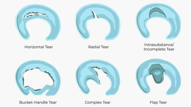
The posterior horn of the medial meniscus is the most common part of the medial meniscus to be injured. A medial meniscus tear typically takes 6-8 weeks to start to recover with physical therapy and appropriate activity modification. In most cases, a meniscus tear can heal on its own provided it is given the right opportunity to recover.
Read our related article to learn more about recovery and exercises.
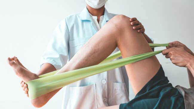
The type of tear, the blood supply to the injured area, and the health of the individual and the tissue will influence healing times. If symptoms persist, the decision may be taken to remove or repair the damaged tissue surgically.
A meniscectomy, where the damaged cartilage is surgically removed, can take 3 to 6 weeks to recover post-surgery. Recovery from surgical repair will be much longer, typically 3 to 6 months. Typically surgery to repair will only be undertaken in individuals and meniscus areas with a good chance of healing.
Typically excessive force through the knee joint with a rotational force, or a sudden acceleration or deceleration will cause a meniscus tear.
Tears of the posterior horn of the medial meniscus are more likely to occur when the knee is in deep flexion, such as squatting or crouching.
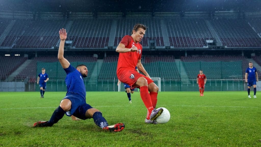
Common sports for acute meniscus injuries are football, rugby, skiing, wrestling, basketball, and baseball. But any sport with high volumes of jumping and running with sudden changes of direction or speed is at risk.
It is common for a meniscus tear to occur with an injury to the ACL.
Other risk factors for degenerative tears include being over the age of 60 years, men being more at risk than women, occupations that require frequent kneeling and squatting, and stair climbing.
This article is written by James McCormack, a Lower Limb Specialist who is an expert in treating Knee Pain.
This is not medical advice. We recommend a consultation with a medical professional such as James McCormack if you are experiencing any of the symptoms discussed in this article. James offers Online Physiotherapy Appointments weekly and face-to-face appointments in his London clinic.