Knee Ligament Injury
LCL Injury
Read More >

Minute Read
|Posted 4 years ago
|Last updated: 19/11/2023
|by James McCormack

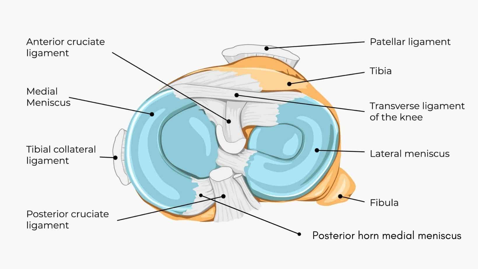
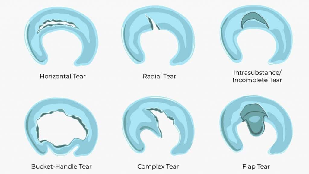
The meniscus is two crescent-shaped, thick pieces of cartilage that sit in the knee between the tibia and the femur. There is a medial and a lateral meniscus. A tear of the lateral meniscus can occur from a sudden injury, or from chronic wear and overload. There are many many different types of tears that can occur, named as a description of the form of tear: horizontal, radial, intrasubstance/incomplete, bucket-handle, complex/degenerative or flap tears. A tear of the lateral meniscus may also be termed related to the location of the tear, this may be the anterior or posterior horn of the lateral meniscus.
Finally, the location of lateral meniscus tears may be defined as related to the blood supply. The meniscus as a whole has a relatively poor blood supply. There is some blood supply to the outer rim of the meniscus, the red area. But no blood supply to the middle portion which is termed the white area.
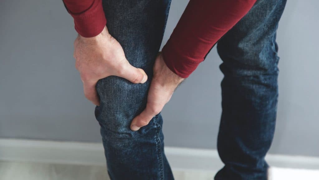
Symptoms of a lateral meniscus tear are varied depending on the severity of the damage to the meniscus as well as many other factors, such as body strength and health, pain sensitivity as well as the psychological impact of the injury. Usually, the pain of a lateral meniscus tear will feel like an ache or sharp sensation at the outside of the knee. However, lateral meniscus tear pain location can move around from the outside, or to the back, or sometimes medial pain is felt at the inside of the knee. There can be calf pain or general pain don’t the leg.
There is often swelling, that is diffuse and can make the knee look a bit puffy or you might notice a loss of definition of the bony prominences or dimples in the knee. The swelling is predominantly in the joint, known as joint effusion, and can make it feel like you can’t bend your knee. This is why flexion is usually limited, and the terminal range of extension may be lost.
With an acute injury pain and swelling may be immediate or may develop up to 48 hours after the injury occurred. The pain usually comes on before swelling. Minor injuries may have no swelling.
If the injury to the lateral meniscus is significant you may feel instability or get a locked knee. Locking of the joint occurs when the tear has a free part that gets folded or stuck in the joint preventing movement. A flap tear or bucket-handle tear, are good examples of this. This will also give the feeling of instability as the joint surface is inconsistent. These free-to-move fragments can also give a sensation, or sometimes an audible, clicking or popping.
As the meniscus has a poor blood supply, bruising is rare with this injury. In most cases, the presence of bruising would indicate that another structure may be injured, separately or in conjunction with the lateral meniscus.
A lateral meniscus tear can be caused by an acute or chronic overload of the meniscus. Acute injuries are often related to a twisting of the knee, an awkward movement when squatting or squatting with heavy weight.
Chronic injuries will be related to repeated positions of stress on the meniscus and may be related to your job, hobbies or habits. For example, if your work involves picking up heavy weight from low positions and you are regularly bending and lifting this can take its toll on the meniscus.
In sports, the mechanism of injury is commonly a twist of the knee when the foot is fixed or hyperextension of the knee, for example, a tackle in football. A rare cause of a tear can result from a Discoid Meniscus.
Lateral meniscus tears are diagnosed based on the reported symptoms and a physical examination by a physical therapist or sports doctor. The physical exam will evaluate your strengths, weaknesses and flexibility, as well as stressing different structures to assess what structure is producing your symptoms of pain.
There are other structures that can cause similar symptoms, so a differential diagnosis list should be made and structures should be tested to rule them in or out as possibilities for your diagnosis. If there is any doubt, radiological imaging will be used to confirm the diagnosis.
The classification of your injury may be made in the clinic, based on your symptoms or with MRI imaging. Grade 1 to 3, have an increasing size of the tear and increasing symptoms.

There are several tests that can be used for stressing the lateral meniscus to assess if there is an injury or tear. McMurray, Thessaly and Apley are all clinical, orthopaedic tests that are used. Each of these tests puts pressure on the meniscus in different positions of knee flexion and adds rotation through the knee. From these, and with clinical symptoms relevant to a meniscus tear, a clinical diagnosis can be made.
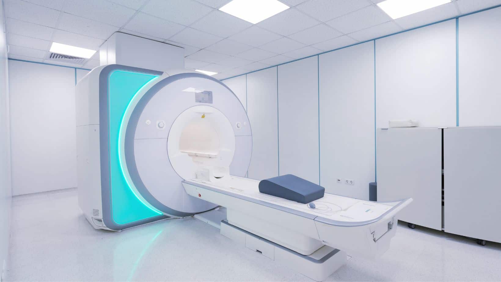
MRI imaging is used along with a clinical examination to diagnose a later meniscus tear. An MRI of a lateral meniscus tear is the most sensitive and accurate form of radiological imaging that can be used. But unfortunately, MRI scans have a high cost.
Another radiology that can be used for diagnosis is ultrasound, An ultrasound image will show if there is a tear in the medial or lateral meniscus with high sensitivity but is not able to specify the type or grade of meniscus tear unline MRI (Mostafa et al, 2019). The cost of ultrasound imaging is much lower, however, and therefore may be more accessible to a greater population.
This is not medical advice. We recommend a consultation with a medical professional such as James McCormack. He offers Online Physiotherapy Appointments for £45.