Knee Exercises
Exercises for Patellar Tendonitis
Read More >

Minute Read
|Posted 4 years ago
|Last updated: 23/11/2023
|by James McCormack

Patella tendonitis is also known as patella tendinopathy or Jumper’s knee. It is a condition of pain in the patellar tendon at the front of the knee. It affects the tendon below the patella, while quadriceps tendonitis affects the tendon above the patella.
The patella tendon attaches the quadriceps muscle to the tibial tuberosity on the shin bone. The patella bone sits within this tendon. When a tendon is overloaded, it changes. We call this a state of disrepair.
In this state, its structure and physiology change. It develops a greater number of new blood vessels and nerve endings and can retain more water. Due to these changes, it becomes less tolerant to load and can have a number of symptoms.
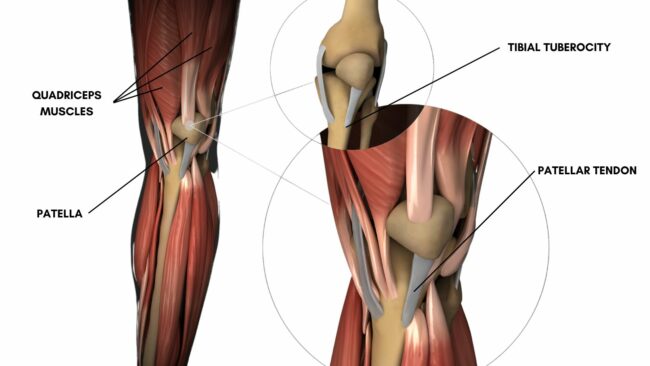
Pain is located at the lower end of the tendon, at the insertion to the tibial tuberosity. Sometimes, it can be felt at the lower end of the patella, where the tendon attaches to the bone also. It can affect one or both knees.
Pain will be described as a dull ache or soreness. It can become sharper and more severe as the condition progresses.
In addition to pain, tightness in the quadriceps muscle and stiffness to bend the knee are common. A click or popping sensation may be felt. Similarly to pain, this usually improves with movement.
Typically, there will be no bruising or swelling but, the tendon can become thickened. This can appear and feel like a lump or bump in the tendon.
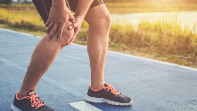
When irritable, the tendon can be painful with any use. It is common for it to hurt to walk, and movements such as descending stairs, squatting, lunging and especially jumping can be very painful. Pinching or compressing the tendon will increase pain, so kneeling or bending the knee fully can also be problematic.

Typically, tendon pain will hurt more at the start of an activity and after periods of rest, and ease as the muscles and tendon warm up.
Pain commonly returns, and may be worse a few hours or the day after too much activity.
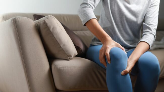
Stiffness will be more significant first thing in the morning or after long periods of being sedentary.
Patella tendonitis is caused by acute or chronic overload. Acute may be a trauma such as a direct impact or fall. Chronic may be repetitive overuse with insufficient recovery or poor conditioning.
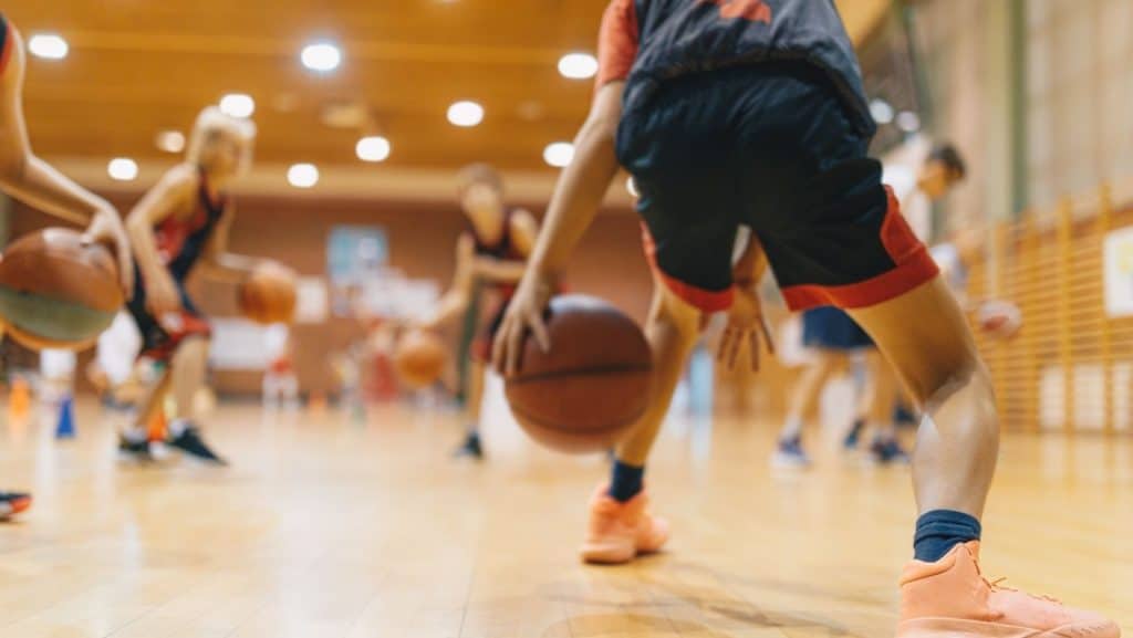
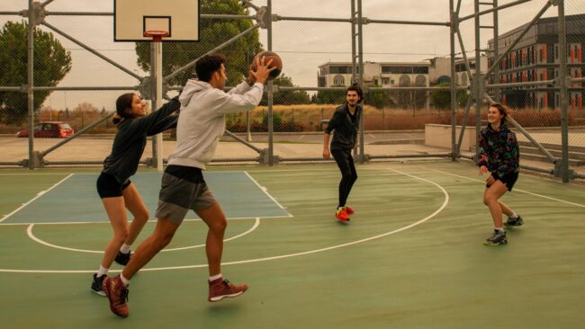
It is common in athletes of jumping sports, such as basketball, netball, gymnastics, and jump rope. It can also be present in people who do not do these sports but do repetitive and powerful knee bending and straightening actions, such as runners.
The patella tendon is also stressed when the knee bends over the foot with weight, such as hiking or trail running, especially going downhill. Tennis players lunging forwards for a ball, weight lifting in the gym, squatting or deadlifting, and many other occasions.
It is more common in the non-athletic population over 35 years old. As we age, our tendons become less flexible and are less able to tolerate stress. Therefore, any changes to training or increase of load to the tendon need to be done more gradually than when younger.
Research suggests that men are more susceptible than women and basketball and netball are the most common sports related to patellar tendonitis.
An experienced physical therapist or sports doctor will discuss your history and symptoms. This will guide their physical examination.
Patellar tendonitis is diagnosed with a cluster of tests and symptom patterns.
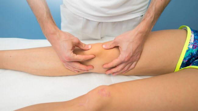
The patellar tendon will be pinched or compressed to see if it reproduces symptoms. Palpation around the tip of the shin bone where the tendon attaches to the tibial tuberosity is usually painful.
Testing the quadriceps to straighten the knee, against resistance, will typically provoke pain. As will some movements such as squatting with knees over toes or fully bending the knee and stretching the quads muscle. If these are pain-free the tendon may need to be loaded more with single-leg squats, or hopping.
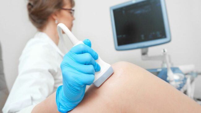
Ultrasound and MRI are valuable tools for looking at the health of the tendon. Both can show tendinopathic changes such as more new blood vessels and nerves. And can also show areas of inflammation, calcification, small tears, or the development of bone spurs.
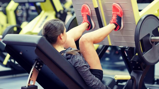
The two most used rehab protocols for Patellar Tendonitis are heavy, slow resistance training and eccentric loading. Both work by strengthening the quadriceps muscle and patellar tendon. The use of a decline board for squatting has also been shown to be an effective way to load the patellar tendon. Another article discusses these in more detail: Exercises for Patellar Tendonitis.
Other exercises can help to facilitate the healing of the patellar tendonitis. Foam rolling and massage, which can be done with a massage gun, help relax the quadriceps muscle. Strengthening surrounding muscles such as the gluteals, calf and hamstring muscles with bridge, clam, and calf raise exercises.
Acupuncture needles can be used directly around the tendon to improve blood supply and to the surrounding muscles to reduce muscle tone and tension. Additional needle points can be used further away from the knee, such as in the hands and feet, for their effect on the sensitivity of the nervous system as a whole.
Corticosteroid injections are effectively used to reduce inflammation and pain short term. However, long-term outcomes, are poor.
Similarly, platelet-rich plasma injections (PRP) are used with variable success but generally are not considered very effective for patellar tendonitis.
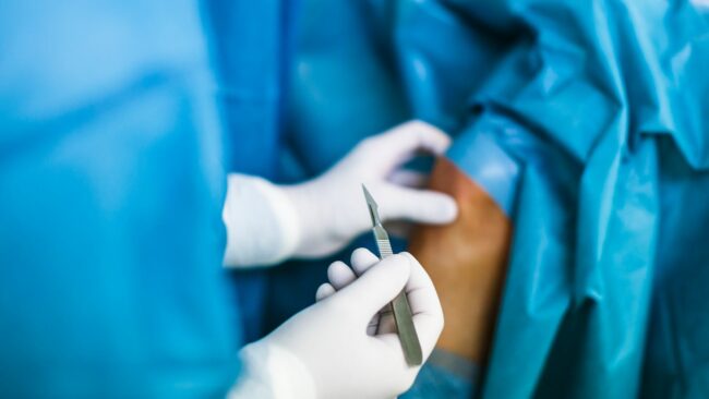
Traditional surgery is used less and less for treating patellar tendonitis. It involves debriding the tendon and cutting out portions of degenerative tissue. Research shows low success rates.
If symptoms have persisted for 3-6 months a tendon scraping procedure can be used. This is much less invasive than surgery. It also has a greater success rate when combined with rehab. Scrapping removes the abnormal nerves that grow on the surface of the tendon and can reduce pain.
This article is written by James McCormack, a Lower Limb Specialist who is an expert in treating tendon and Knee Pain.
This is not medical advice. We recommend a consultation with a medical professional such as James McCormack if you are experiencing any of the symptoms discussed in this article. James offers Online Physiotherapy Appointments weekly and face-to-face appointments in his London clinic.