
Ankle Ligament Injury
Sprained Ankle
Read More >

Minute Read
|Posted 4 years ago
|Last updated: 19/03/2023
|by James McCormack

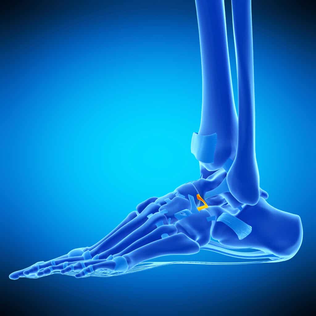
A ligament connects a bone to a bone, and the Bifurcate ligament is Y-shaped with two bands: the calcaneonavicular ligament (CNL) and calcaneocuboid ligament (CCL) As the foot moves into inversion, it increases stress on the Bifurcate Ligament; if this is greater than the ligament can tolerate, it can result in a tear or Avulsion fracture.
A study of 53 cadavers found that 90.6% of feet had both (CNL) & (CCL). 9.4% had the CNL and no CCL, and none (0%) had the CCL and no CNL.
Bifurcate Ligament Sprain symptoms include pain on the inner aspect of the foot close to the heel bone, and there may be some subtle swelling on the hindfoot.
Weight-bearing in standing or walking can be painful, as can plantarflexion or supination of the foot.

Lateral ankle sprains account for 85% of all ankle sprains and it is the most common cause of a Bifurcate Ligament injury as it places high force and strain on the ligament.
The likelihood of a Bifurcate Ligament Sprain increases if the ankle sprain occurs while the foot is in plantarflexion which is common for people wearing heels.
The origin of the Bifurcate ligament is the Anterior Process of the Calcaneus, and this is a common site of avulsion fracture to the Bifurcate Ligament post-ankle sprain.
A clinical assessment can reveal slightly distal pain to the specific location of a lateral ankle sprain where there may be pain on palpation of the Anterior Process of the Calcaneus.
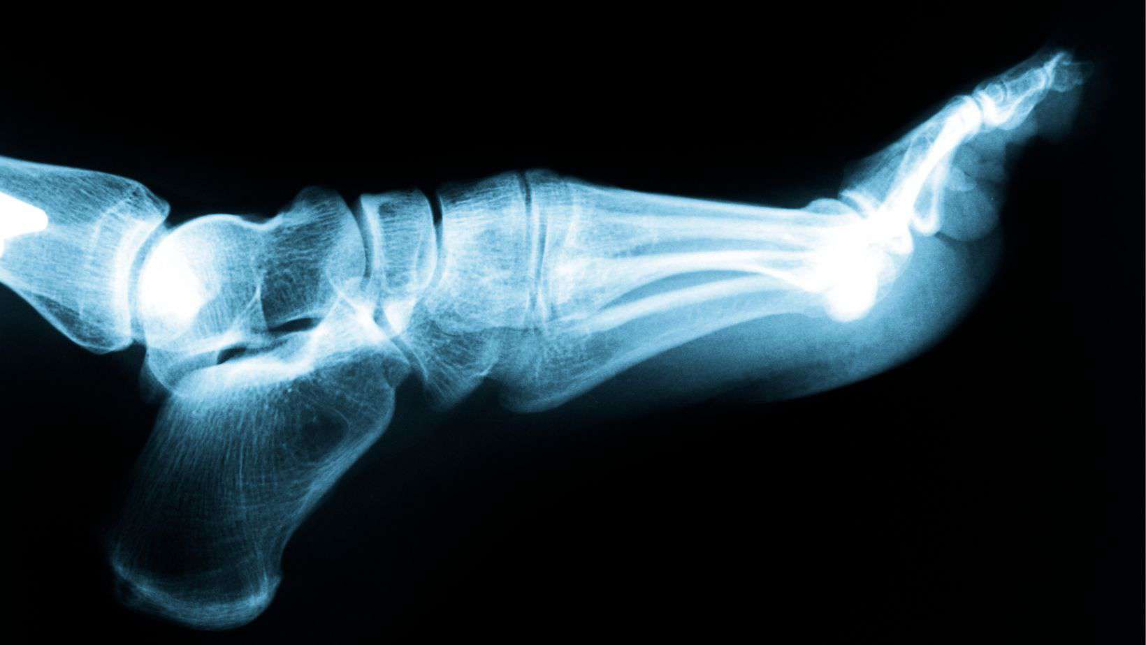
An x-ray is the most common scan for an ankle sprain and an oblique x-ray can identify an Avulsion fracture of the Bifurcate Ligament.
An MRI can establish greater clarity over a Bifurcate Ligament Sprain, while a CT Scan can provide more information on an Avulsion fracture.
Treatment for a Bifurcate Ligament avulsion fracture is normally conservative. It commences with 4-6 weeks in a walker boot, where a patient weans out under a physical therapist’s guidance as pain allows.
Strengthening and balance exercises can address muscular atrophy from wearing the boot, and an ankle brace may be recommended for extra support in the early stages of rehabilitation.
As the foot and ankle strengthen, rehabilitation should become more dynamic with plyometric and change of direction exercises. The total time for recovery from a Bifurcate Ligament avulsion fracture is normally up to 12 weeks.
We have written a separate article on common questions about playing various sports while recovering from ankle sprains.
Unstable or comminuted fractures may need surgery, which should be discussed with an Orthopaedic consultant.
This is not medical advice and we recommend a consultation with a medical professional such as James McCormack before trying any of these exercises. James offers Online Physiotherapy Appointments