
Foot Bone or Joint Injury
Accessory Navicular (Os Naviculare)
Read More >

Minute Read
|Posted 4 years ago
|Last updated: 25/01/2024
|by James McCormack

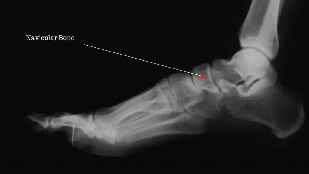
The navicular bone is located on the inner aspect of the medial arch of the foot; it is saddle-shaped and sits between the Talus bone and the 3 Cuneiform bones. The Navicular bone plays a fundamental role in the transfer of weight and forces from the hindfoot through to the mid and forefoot while also providing structural stability to the medial arch of the foot. The blood supply of the Navicular bone plays a vital role in its vulnerability when recovering from stress fractures.
Two arteries enter the bone centrally but immediately diverge medially and laterally, leaving the central aspect of the bone with low blood supply levels. Repeated stress to the central aspect of the Navicular Bone from impact sports can cause a Navicular Stress Fracture.
When describing the symptoms of a Navicular Stress Fracture, patients often report a slow, gradual onset of dull pain on the medial arch of the foot that is worse with activity but improves with rest. There may be some mild swelling or bruising around the navicular bone, which is often not present.
The location of the pain in patients is approx 19mm in diameter around the top of the navicular, closer to the ankle bone, and this is known as the N-spot. It has been found that 81% of patients have pain on palpation of the N-spot.
Due to the unique position of the Navicular bone, high forces are anatomically placed through the central aspect of the Navicular when walking and running; this is coupled with strain placed on the Navicular from the Posterior Tibial Tendon. When impact activities such as running and jumping and repetitively carried out, these forces can cause a Navicular Stress Fracture.
As a result, most Navicular Stress fractures are found in athletes. In a study of 200 individuals with Navicular Stress Fractures, most were males, and 98.5% were athletes.
A clinician such as a Physical Therapist can identify the likelihood of a Navicular stress fracture following a physical examination that involves pain on palpation of the N-Spot, pain on single leg calf raise and a single leg hop test.
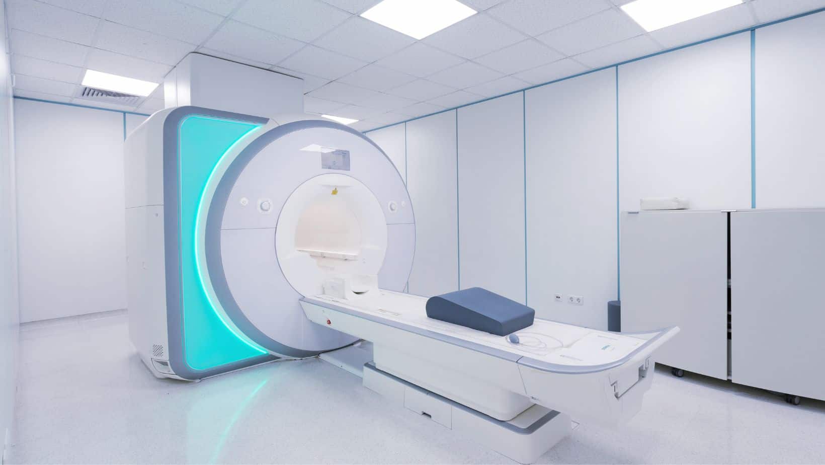
A standing x-ray can detect a Navicular Stress Fracture, but this is difficult in the early stages of injury, so an MRI is preferred if possible. The gold standard form of imaging for a Navicular Stress Fracture is a CT-Scan, which is often difficult to source.
If a stress fracture is found, checking your Vitamin D levels with or without a Dexa Scan is often recommended to check your bone health.
Early detection of this injury is vital, and a consultation with your Physical Therapist and Orthopaedic consultant on the best treatment is recommended. In non-operative cases, patients are usually placed in a cast or a boot for 6-8 weeks, non-weight bearing before increasing weight through the joint and eventually weaning out of the boot.
Mass atrophy of the foot and ankle will result from the time spent in the boot, and your Physical Therapist should develop a specific strengthening, mobility and stability rehabilitation protocol. A rehabilitation protocol should be progressively difficult, pain-free, and more sports-specific over time. A gait analysis can provide information on the most appropriate footwear and the necessity for custom insoles.
If a patient has low vitamin-D levels, it can influence the bone healing rate, increasing the time spent in a boot and the speed that a patient works through a rehabilitation protocol.
There are a number of surgical techniques that can be carried out for a Navicular Stress Fracture, and these should be discussed with an Orthopaedic Consultant.
This is not medical advice. We recommend a consultation with a medical professional such as James McCormack. He offers Online Physiotherapy Appointments.
Related Article: Accessory Navicular Syndrome – Kohler’s Disease