Foot Exercises
Accessory Navicular Syndrome Exercises
Read More >

Minute Read
|Posted 4 years ago
|Last updated: 17/09/2023
|by James McCormack

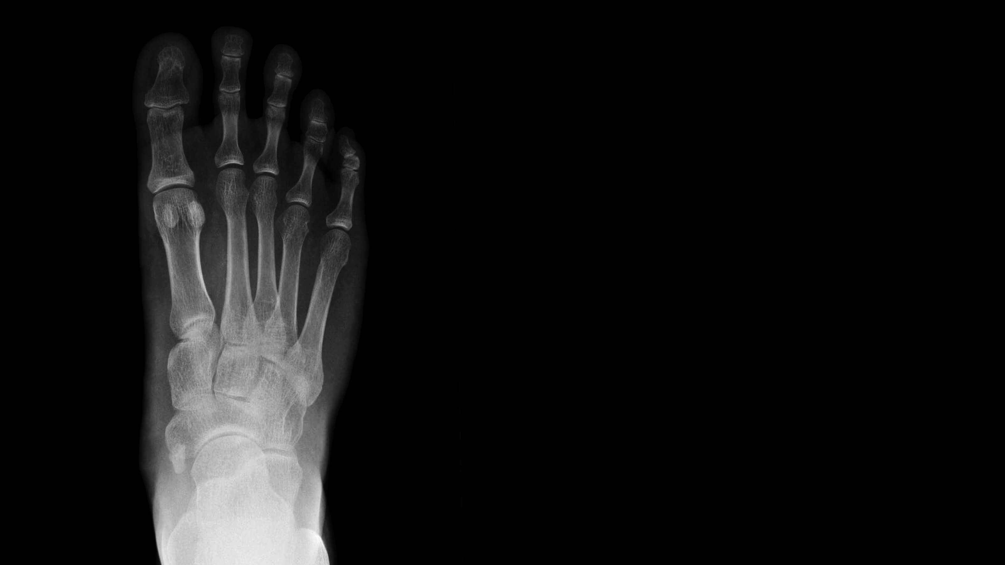
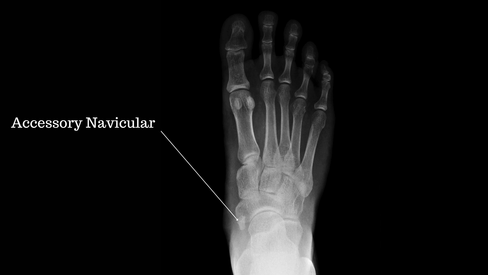
The navicular bone is a wedge-shaped bone that sits in the foot’s medial arch between the talus and 3 cuneiform bones playing a vital role in the structural integrity of the foot’s arch.
The navicular bone plays a vital role in the stability of the medial column of the foot, and due to its numerous attachments from tendons and ligaments, it plays a fundamental role in the gait cycle.
There are two ossification points on the navicular bone (where cartilage turns to bone) during childhood; in a low proportion of the population, these points do not fuse. In these cases, a small piece of additional bone or cartilage remains, known as the accessory navicular bone or Os Naviculare.
One study on 266 asymptomatic Koreans found a prevalence of accessory bones of 49.2%; of those, an accessory navicular accounted for 34%. Another cross-population study found that accessory naviculars occur more often unilaterally with equal distribution between males and females.
When the accessory navicular bone becomes irritated, it is called painful Accessory Navicular Syndrome.
The symptoms of accessory navicular syndrome are pain along the medial arch of the foot, specifically over the navicular bone. There may be some mild localised swelling in the area, and it is tender to palpate the navicular bone with a visual bony prominence on the foot’s arch.
Patients may notice friction or tenderness along the arch in tight-fitting shoes, and wearing high-heeled shoes may be uncomfortable.
It can be painful when walking, especially on uneven surfaces while high-impact activities such as hopping or running can significantly irritate the symptoms. Standing for long periods in unsupportive footwear can increase pain levels and it is not uncommon to feel a throbbing sensation in the foot after use.

Those with flat feet appear to have a higher prevalence of accessory naviculars, although little evidence indicates that flat feet cause an accessory navicular.
Tight or ill-fitting footwear can lead to excess friction on the Os Naviculare, potentially negatively affecting the ossification site.
Repetitive foot overuse can cause navicular pain syndrome and a history of ankle sprains.
Ethnicity does play a role in this condition; accessory navicular syndrome is prevalent in approximately 10% of the general public, while there is a prevalence of up to 46% in the Asian Population.
If you have the symptoms of Accessory Navicular syndrome, your clinician will likely refer you for an x-ray in the initial instance, as this is an easily accessible and cheap form of imaging.
If the diagnosis is confirmed on an x-ray, you may be referred for an MRI or Ultrasound scan to identify any swelling or other soft tissue injuries.
CT scans are rarely used and preferred for exact measurement or grading of fractures to the navicular.
A Physical Therapist is best positioned to provide treatment for Accessory Navicular syndrome. Treatments such as taping, joint mobilization and massage can relieve pain alongside home treatments such as resting, anti-inflammatories and icing.
A physical examination will identify muscular imbalances and weakness that can contribute to your pain, and a comprehensive programme of strengthening, balance and stretching exercises will be formed.
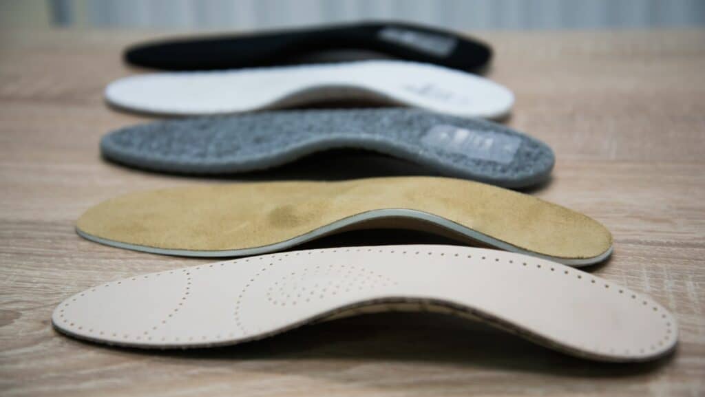
Footwear advice is essential as the correct shoe can relieve pain alongside insoles and orthotics.
A steroid injection may be required for symptoms that fail to settle with Physical Therapy, usually in the form of an ultrasound-guided corticosteroid injection. Depending on the consultant, 1-2 weeks in a walking boot may be recommended before recommencing Physical Therapy.
In a few cases, conservative treatment is unsuccessful, and surgery is required. Open excision of the accessory navicular is the most common form of surgery for this condition, which has a high success rate.
This is not medical advice. We recommend a consultation with a medical professional such as James McCormack if you have foot pain. He offers Online Physiotherapy Appointments weekly.