Braces and Supports
LCL Knee Brace
Read More >

Minute Read
|Posted 4 years ago
|Last updated: 07/12/2023
|by James McCormack

The LCL is the lateral collateral ligament. A ligament is a thick strong band of connective tissue, that attaches one bone to another. Their function is to keep the bones together but allow for movement in some directions while preventing movement in others. Here we will discus the LCL of the knee, you can also find an LCL at the elbow.
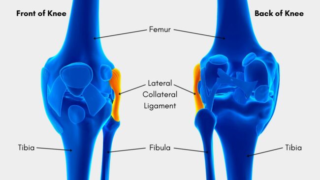
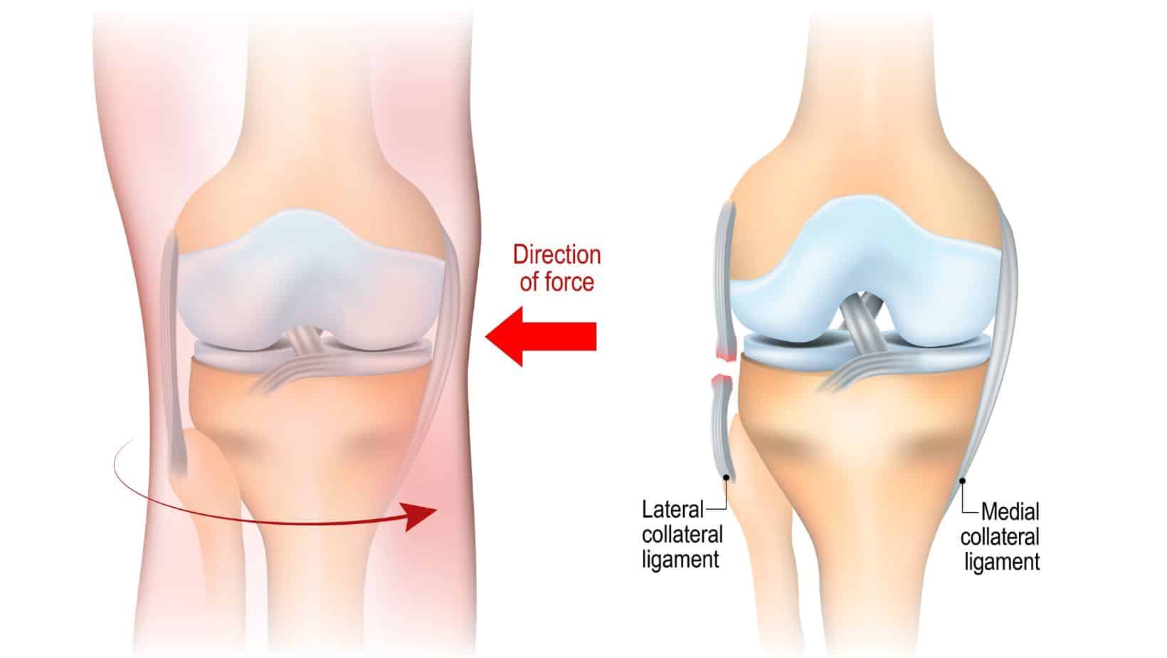
The LCL is on the outside of the knee, connecting the femur and fibular bones, and also attaches to the iliotibial band.
The main function is to provide lateral stability to the knee by preventing varus movement. In addition, it can prevent rotation of the knee and hyperextension. This action is particularly important if the cruciate ligaments are injured.
Injury to the lateral collateral ligament can range from minor sprains to complete tears. Symptoms of an LCL sprain include:

Most LCL sprains are caused by impact or a force to the inside of the knee, pushing it outwards. It is an uncommon injury of the knee, 1.1% of knee injuries in an athletic population.
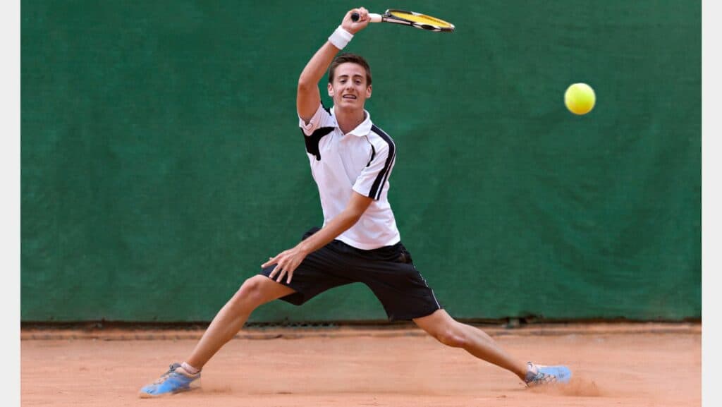
Due to the high force needed to injure this ligament, it is more commonly seen in the younger athletic population. It is more common with tennis and gymnastics. But also with football, rugby, skiing and wrestling sports such as jiu-jitsu.
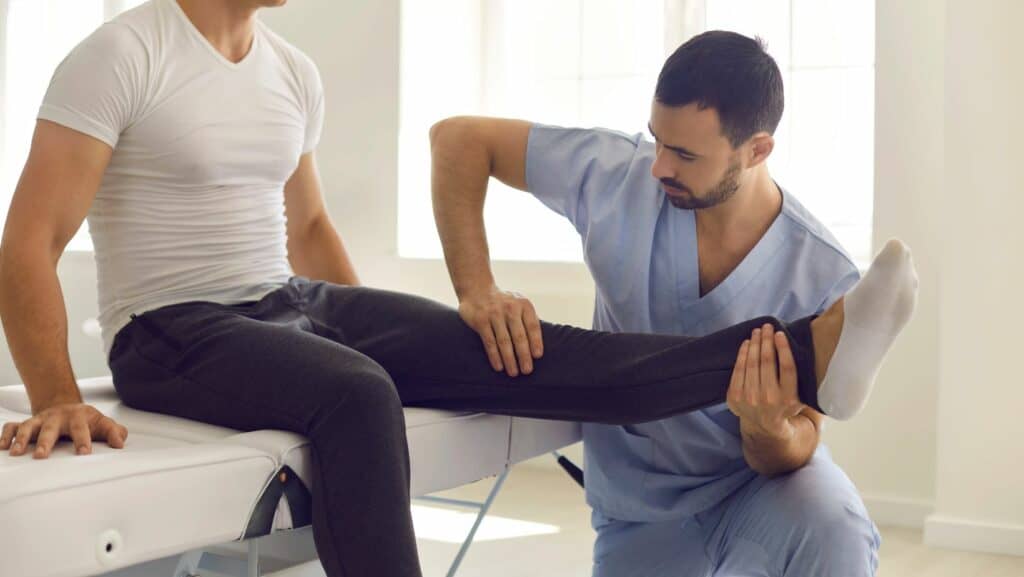
Assessment of a suspected LCL injury is best done by an experienced physical therapist or sports doctor.
There are several other injuries that can present with pain and swelling to the outside of the knee, such as iliotibial band syndrome or a lateral meniscus injury.
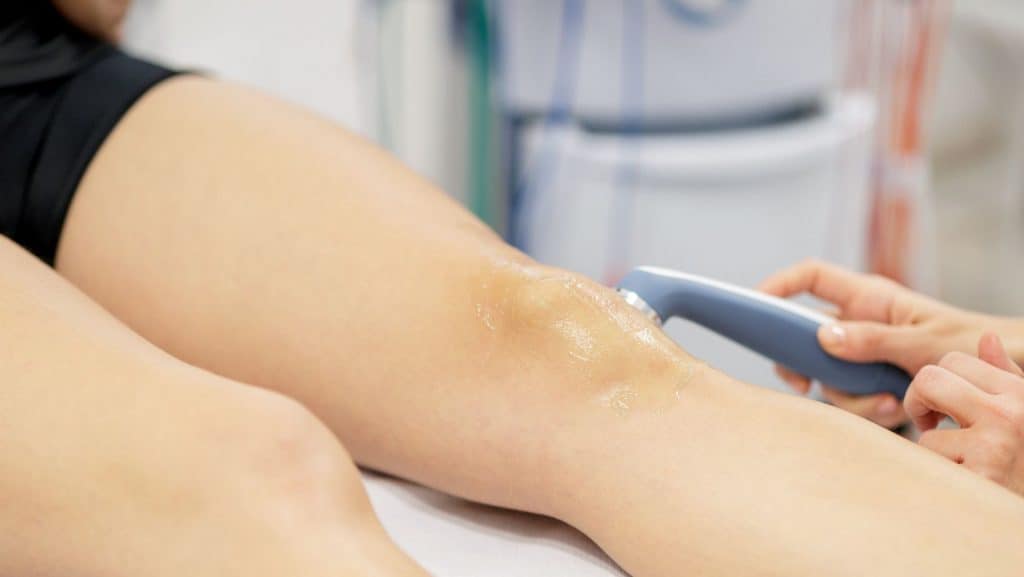
Your doctor or physical therapist will listen to the mechanism of injury and the symptoms you have experienced to direct a physical examination. Diagnosis may be confirmed with radiological imaging such as MRI or ultrasound.
One of the important tests to be included during a physical examination is the adductor or varus stress test. A positive test will reproduce the patient’s pain. The clinician may also be able to feel laxity of the joint, an excess varus movement, which can help to grade the injury.
Minor ligament damage with no laxity.
Symptoms: pain, minor swelling, minor bruising if any and no instability.
Treatment: rest for several days, no sports, reduced walking and elevate leg for swelling. In a minority of cases, crutches might be offered to help mobility in the first week or two if pain is high with movement.
Moderate ligament damage with laxity.
Symptoms: pain, significant swelling, significant bruising and some instability.
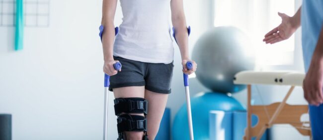
Treatment: will require complete rest for several weeks and elevation of the leg for swelling. A period of 2-4 weeks of using crutches and/or wearing a hinged brace, to completely offload the joint.
Severe ligament damage or complete rupture, significant laxity.
Symptoms: pain, extensive swelling, extensive bruising and instability and loss of varus control.
Treatment: will require a long period of complete rest and elevation of the leg for swelling. A brace and crutches will be necessary for 6 weeks or more. Some will need surgery, for the reconstruction of the injured ligament.
The acute treatment for LCL sprains involves relative rest, ice, compression and elevation. The duration of this will depend on the grade of the injury.
Following the initial phase of rest, active treatment can be started. The most effective recovery comes from rehabilitation exercises. A physical therapist can assess you to prescribe you a specific programme to follow. You can read about the commonly prescribed exercises in our related article: LCL Injury Exercises.
LCL sprain recovery time depends on the grade of injury and an individual’s health and fitness. Grade 1 injuries may take about 3-4 weeks, grade 2 will take longer, about 8-12 weeks and grade 3 will take more than 12 weeks and might need surgery.
Elasticated tapes such as Kinesio tape can be effective for pain relief of lateral knee pain from many causes such as an LCL injury. This tape improves proprioception of the joint. This video shows how to tape the knee with KT tape.
Rigid taping can be applied to improve the stability of the knee and prevent the outward movement (varus) that lengthens and strains the LCL. This taping is often used after an injury to prevent further stress to the ligament on return to sport.
If an injury is severe such as a grade 3 sprain or complete rupture, and not recovering with conservative treatment, surgery may be considered. An operation to reconstruct the ligament can improve stability and function of the knee, and is more effective with higher-grade injuries. Success after surgery will be significantly improved with good rehabilitation and strengthening exercises. These should be guided by an experienced physical therapist.
This article is written by James McCormack, a Lower Limb Specialist who is an expert in treating Knee Pain.
This is not medical advice. We recommend a consultation with a medical professional such as James McCormack if you are experiencing any of the symptoms discussed in this article. James offers Online Physiotherapy Appointments weekly and face-to-face appointments in his London clinic.