

Read More >

Director at Flawless Physio
James McCormack is a Knee, Foot and Ankle Specialist Physiotherapist with over 10 years experience.
Alongside his clinical work he has created this website to provide free-to-read content for it's users.
Alongside his clinical work he has created this website to provide free-to-read content for it's users.
Latest posts by James McCormack (see all)
- Anterior Ankle Impingement - July 24, 2022
- Tarsal Coalition - October 12, 2022
- Sural Nerve Pain - October 3, 2022
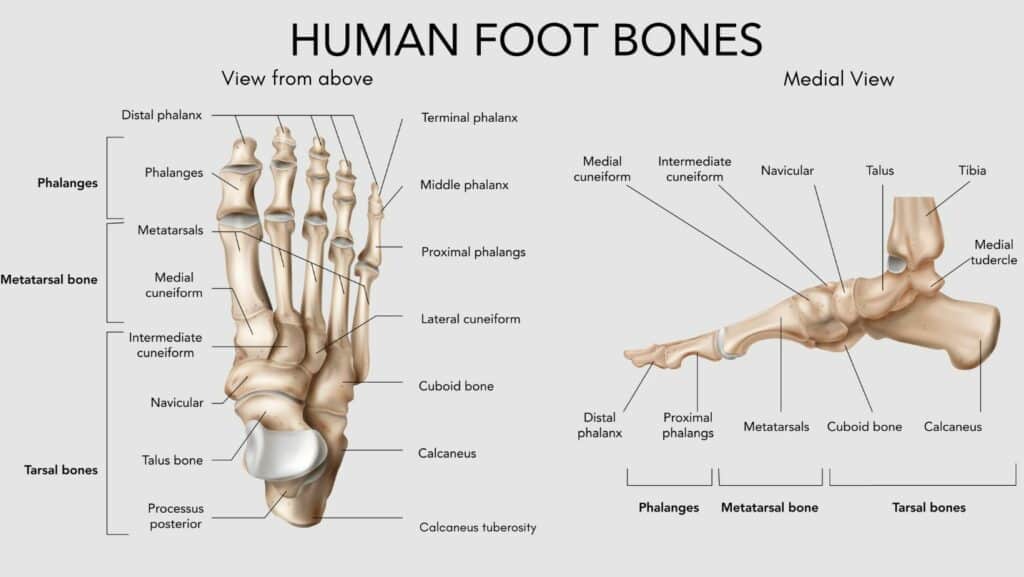
How many bones are in the foot?
There are 26 bones in the foot and 33 joints in the foot. The foot is split anatomically into 3 sections; the hindfoot, the midfoot, and the forefoot. This article will describe in detail the anatomy and function of the major bones in the foot.
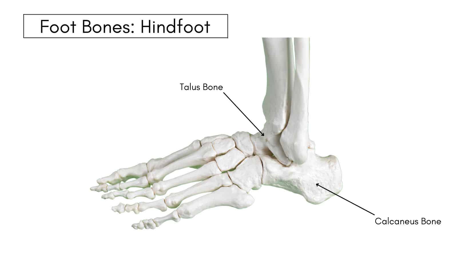
Foot Bones: Hindfoot
The hindfoot consists of 2 bones: the Calcaneus and the Talus bone. Their articulation allows for inversion and eversion of the foot and ankle.
The Talus bone is one of the few bones in the body not connected by ligaments to other bones, and almost its entire surface is covered by cartilage. Its primary function is to transfer force through the heel and forms part of the ankle joint.
The Calcaneus bone is also known as the heel bone and serves as a connection point for the Achilles Tendon and the Plantar Fascia. It is a very strong weight-bearing bone and takes a high-force incident like a fall or car crash to break.
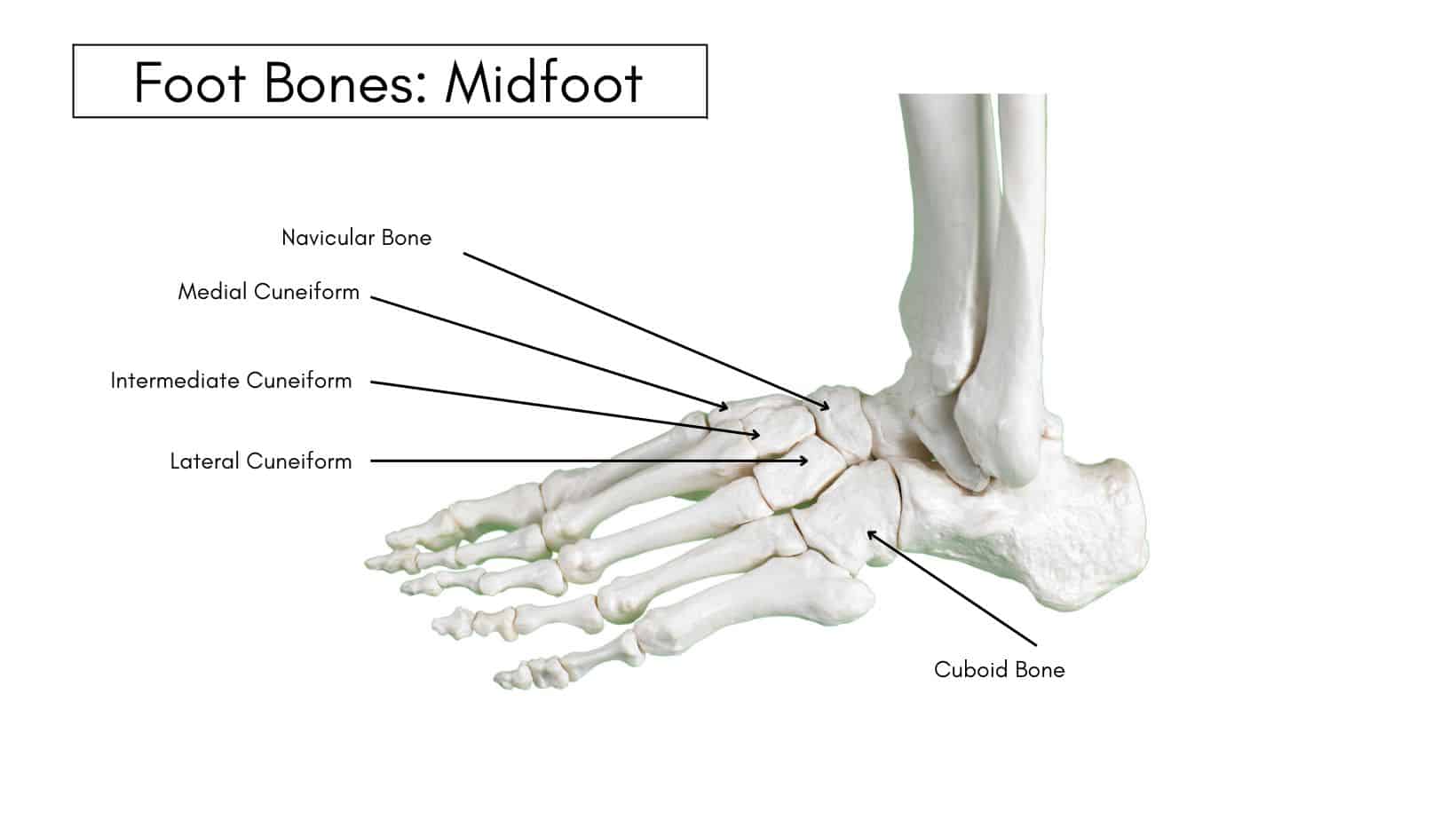
Foot Bones: Midfoot
The Midfoot consists of 5 bones: the Navicular, Cuboid, Medial, Intermediate, and Lateral Cuneiforms. The bones of the midfoot help to form the arch of the foot, and their function is to transmit and reduce forces while allowing the foot to accommodate uneven ground surfaces.
The Navicular bone is irregular in shape and referred to as the keystone of the medial arch of the foot. Due to the location of the Navicular at the peak of the arch of the foot, it is susceptible to stress fractures and can be poor at healing due to its limited blood supply.
The Cuboid bone is roughly cubical in structure hence its name, and it is located on the outside of the midfoot. It helps to provide stability to the midfoot and acts as a pulley for essential structures in the foot.
The Cuneiform bones are 3 wedge-shaped bones within the midfoot. The function of the cuneiform bones is to help form the transverse arch of the foot. They have numerous muscular attachments, such as the Tibialis Anterior, Peroneal Longus, Posterior Tibial Tendon, and Flexor Hallucis Brevis.
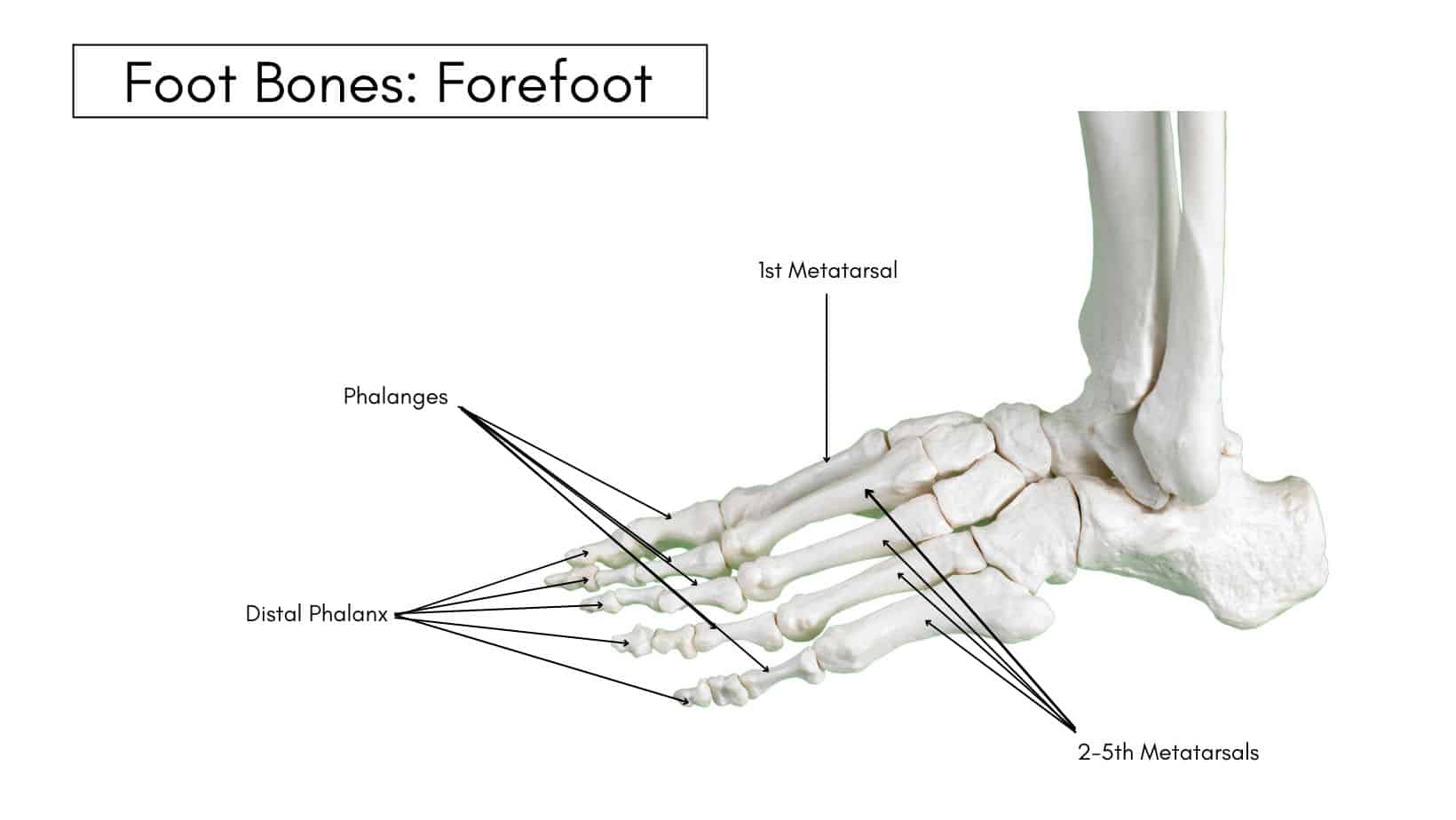
Foot Bones: Forefoot
The forefoot consists of 19 bones; 5 metatarsal bones and 14 phalanges. The big toe has 2 phalanges bones, while the remaining four have 3 phalanges each.
The 1st metatarsal is the shortest and thickest of the metatarsals, and it is designed to take up to 40% of your body weight in standing, which rises to 70% when walking.
The 2nd metatarsal is often the longest, with the 3-5th metatarsals becoming shorter than the other, but this varies depending on foot type, and they assist with balance when walking and running.
The Phalanges provide attachment points to allow the toes to bend and extend while also assisting with balance.
Physiotherapy with James McCormack
This is not medical advice. We recommend a consultation with a medical professional such as James McCormack. He offers Online Physiotherapy Appointments for £45.
Related Articles:
Tendons of the Ankle – Causes of Pinky Toe Pain – Bruised Heel – Foot Pain Chart – Capsulitis of the Foot