Knee Bone or Joint Injury
Tibial Plateau Fracture
Read More >

Minute Read
|Posted 4 years ago
|by James McCormack

The femur is the longest bone in the body. It articulates with the shin bone to make the tibiofemoral joint, which is better known as the knee. The femur has another articulation with the patella, called the patellofemoral joint. The tibiofemoral joint is the largest weight-bearing joint in the body and takes large force when the joint is used in activities such as walking, running, and jumping. The femur is a long bone that widens at its distal end, these flared parts are called the medial and lateral condyles.
Fracture of femoral condyle can occur, although it is a rare injury. It accounts for only about 5% of fracture to the femur, and that is less than 0.5% of all fractures. Fractures of the femur are more commonly at the top, at the neck of the femur, or in the main shaft. The femoral condyles are the lower part of the femur where the shaft widens to two condyles, one medial and one lateral. Both can sustain an injury and become fractured. These fractures are called high-energy injuries due to the high forces needed to cause a break in this strong bone. Typically these injuries are related to a fall from a height or a road traffic incident.
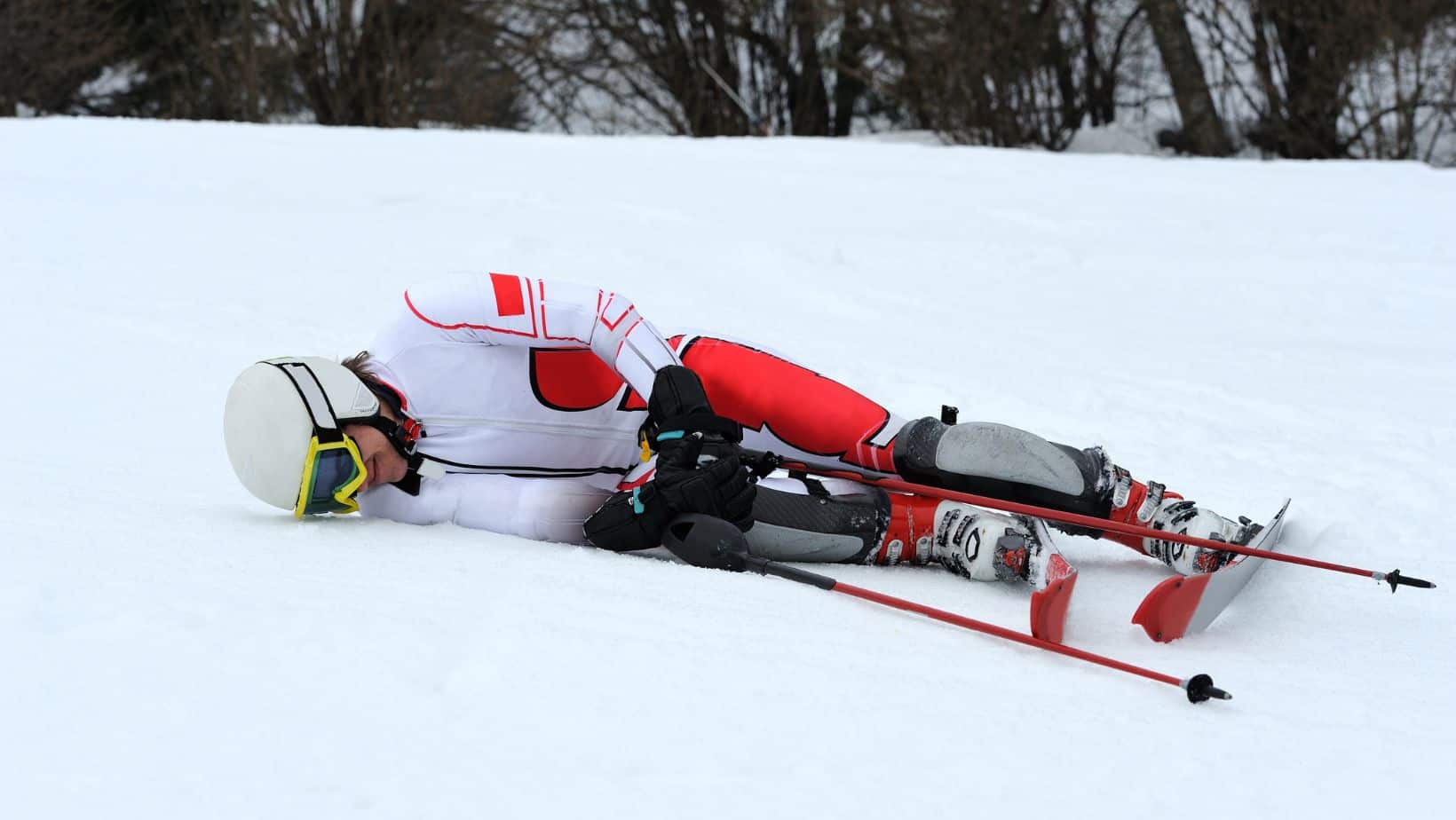
Symptoms are similar to those of any fracture. There will be a sudden onset of severe pain, and inability to weight bear on that leg. Swelling can occur and bruising in many cases. Dependant on the injury the fracture may be close, meaning the skin is not broken or, open where the bone protrudes through the skin.
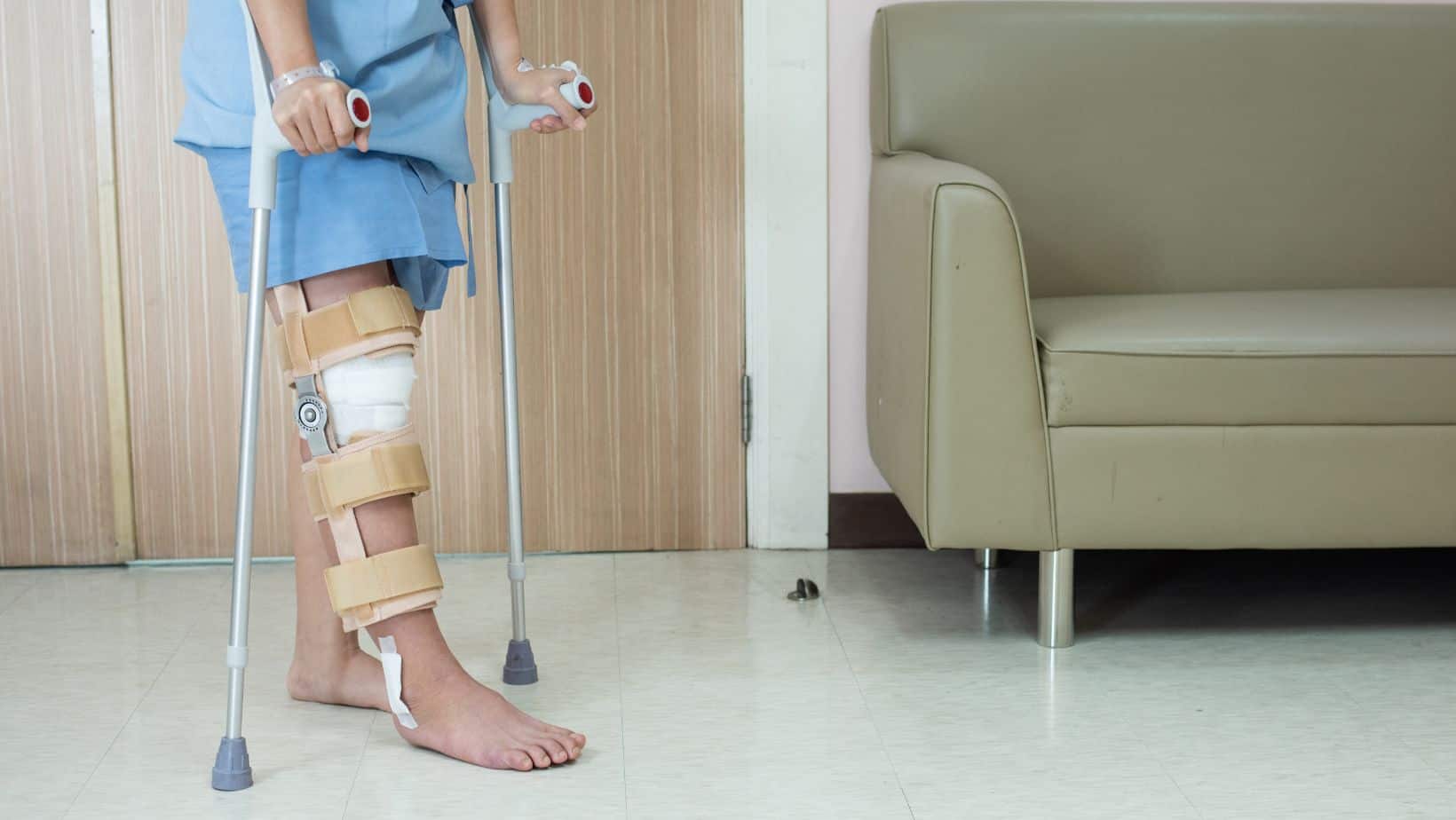
Immediate treatment will need to be at the emergency room. Diagnostic imaging will be necessary and acute treatment of rest, ice, medication and in some cases surgery. Further treatment of these fractures varies dependant on the specifics of the fracture and the other injuries that my have occurred to the surrounding tissues, as well as the individuals health and fitness.
Typically treatment will include rest and time for the bone to heal, this may need surgical intervention followed by a brace of case or may be conservatively manage with a brace or cast. Following this period of healing, knee range of movement will need to be recovered as it will have reduced due to immobility of the joint. Strength in the leg will also need to be regained as this will have also reduced with the inactivity. The longer the inactivity and immobility the longer the recovery and rehabilitation is likely to take.
These are fractures that occur in the coronal plane rather than the more common sagital plane. The coronal plane a vertical plane that runs from side to side and divides the body from front to back. The sagital plane is a vertical plane that runs from front to back and decides the body from side to side. This mean that a Hoffa fracture will be seen on X-ray or MRI from a side view. These fractures account for approximately 40% of all femoral condylar fracture injuries. As it is a high-energy injury it will often be seen with other injuries of the knee.
Hoffa fractures can be of one condyle or can be bicondylar, and are categorised as type 1,2 and 3 depending on the angle of the fracture line, and with letter a,b and c, denoting the region of the femoral condyle that the fracture is in. Types 1 and 3 have a better prognosis due to the location of attachment of soft tissues and blood supply (Zhou et al, 2019).
This is not medical advice. We recommend a consultation with a medical professional such as James McCormack. He offers Online Physiotherapy Appointments.
Related Articles:
Knee Pain Location Chart – Muscles of the Knee – Hoffa Fat Pad