Knee Bone or Joint Injury
How to Treat a Baker’s Cyst Behind the Knee?
Read More >

Minute Read
|Posted 4 years ago
|Last updated: 08/01/2023
|by James McCormack

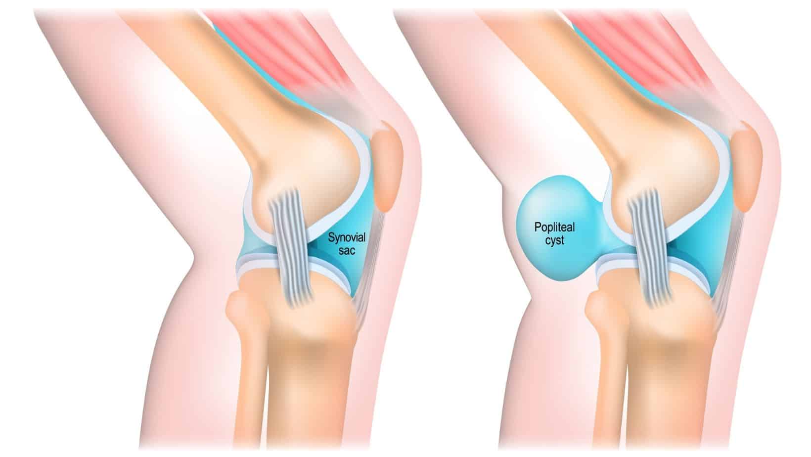
A Baker’s cyst is also known as a popliteal cyst. It is located a the back of the knee in what is called the popliteal fossa. A Baker’s cyst is a collection of fluid that flows out of the knee and forms a swelling at the back of the knee joint. This is an extremely common disorder of the knee, Dr. William Morrant Baker first described the pathology in the 1800s. This condition can affect people of any age, including children, but is most common between the ages of 30-70 years. It can be found in 4-7% of asymptomatic people (usually a small Baker’s cyst), but is more common to occur with symptoms, with a higher prevalence with older age and knee injury or osteoarthritis (Li, 2017).
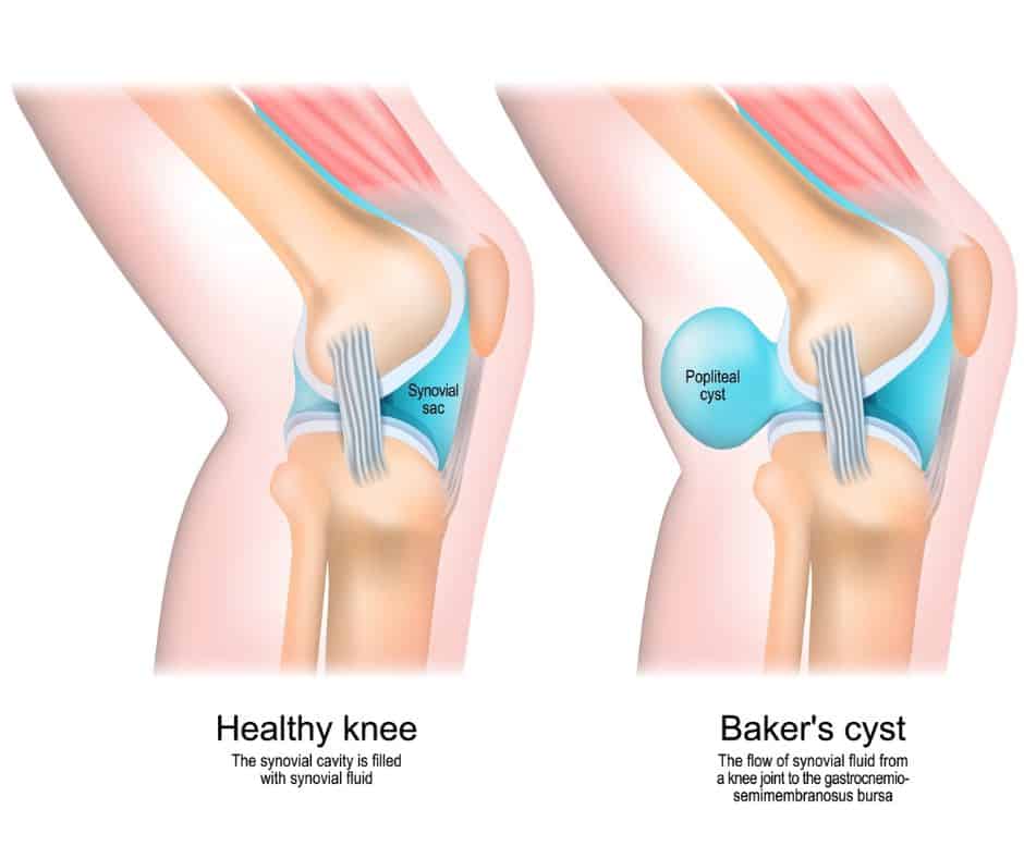
In the majority of cases a Baker’s cyst is painful. The fluid that flows from the joint to form the cyst is inflammatory liquid. This liquid can put pressure on sensitive structures and cause pain at the back of the knee. It is commonly reported that pain is worse when bending the knee, especially when weight bearing. Movement such as squatting are particularly painful as the cyst is compressed under weight. The compression of the cyst will make the knee feel stiff and blocked, usually the full range of flexion at the knee will be limited. The extent to which the knee range of movement is limited is related to the size of the Bakers cyst as well as the pain intensity.
Complications to a Bakers cyst are that they can rupture or become infected. An infected Baker’s cyst can be a serious health issue and will need urgent medical attention as it can lead to sepsis which is life threatening.
The average size of a Baker’s cyst is around 3cm in diameter. The size of the cyst can change related to the amount of synovial fluid that is within it. If the joint has been used and stressed, it is common for the cyst to be larger. Similarly, the size of the cyst will reduce with rest, compression and elevation. There are unusual cases are very large baker’s cysts such as described in this case report, of a baker’s cyst measuring 95mm x 26mm (Adiyeke et al, 2017).
Baker’s cyst causes are commonly related to overuse or degenerative changes in the knee joint. Typically a Baker’s cyst will occurs as a result of knee pathology, most commonly meniscal tears and osteoarthritis. A flair up will be caused by over loading the joint with activity such as walking, running or repeated knee flexion such as squatting. Less commonly, inflammatory arthritis can cause a Baker’s cyst to develop.

A ruptured Baker’s cyst is caused by overuse of the joint once a cyst has developed. Wearing well supporting, cushioned footwear, avoiding excessive exercise and impact, and avoiding knee rotation and repeated flexion are effective ways to reduce the risk of rupture.

A clinical examination in most cases will give sufficient information to diagnose a Baker’s cyst. However, in some cases confirmation may be needed if symptoms are not clear or if symptoms are present that might be caused by alternative diagnosis.
MRI and ultrasound are effective imaging to confirm diagnosis and exclude other diagnosis. A Baker’s cyst on ultrasound will show clearly as excess fluid a the back of the knee in a cyst and fluid between the tendon of semimembranosus and medial head of gastrocnemius muscle. X-ray can be used to rule out other pathologies such a fractures, and can show reduced joint space, suggestive of osteoarthritis, but is much less sensitive, so would therefore not confirm a diagnosis of a Baker’s cyst.
A ruptured Baker’s cyst symptoms are very similar to a DVT, including a painful, swollen, tight and red calf. A crescent sign is a bruise like discolouration in the shape of a crescent moon at the inside of the ankle, below the medial malleolus. It is a differential sign suggesting a ruptured Baker’s cyst, rather than a DVT. It is suggested to be the only indicator from a clinical examination to differentiate between the two (Mizumoto, 2019). Colour Dopplar can be used with ultrasound to rule out vascular pathology such as DVT.
Management of a Baker’s cyst primarily involves rest, compression and elevation to help drain the fluid from the cyst to reduce pain and improve range of movement. If the Baker’s cyst is very painful, aspiration of the fluid to reduce the size and pressure and a corticosteroid injection to reduce inflammation can be effective. You can read more about how to treat a Baker’s cyst in our related article: How to Treat a Baker’s Cyst Behind the Knee?
This is not medical advice. We recommend a consultation with a medical professional such as James McCormack. He offers Online Physiotherapy Appointments for £45.