Braces and Supports
Jumper’s Knee Brace
Read More >

Minute Read
|Posted 4 years ago
|Last updated: 19/11/2023
|by James McCormack

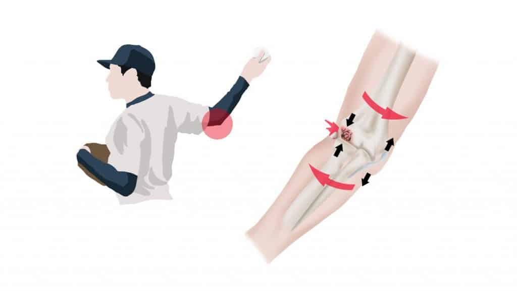
Osteochondritis dissecans is a condition that can spontaneously occur in any joint between the bone (osteo) and cartilage (chondral) caused by a loss of blood flow to these tissues. The specific cause of the restricted blood flow is not fully understood but OCD is linked to trauma to the joint, periods of physical activity causing micro trauma to the joint, and some genetic predispositions.
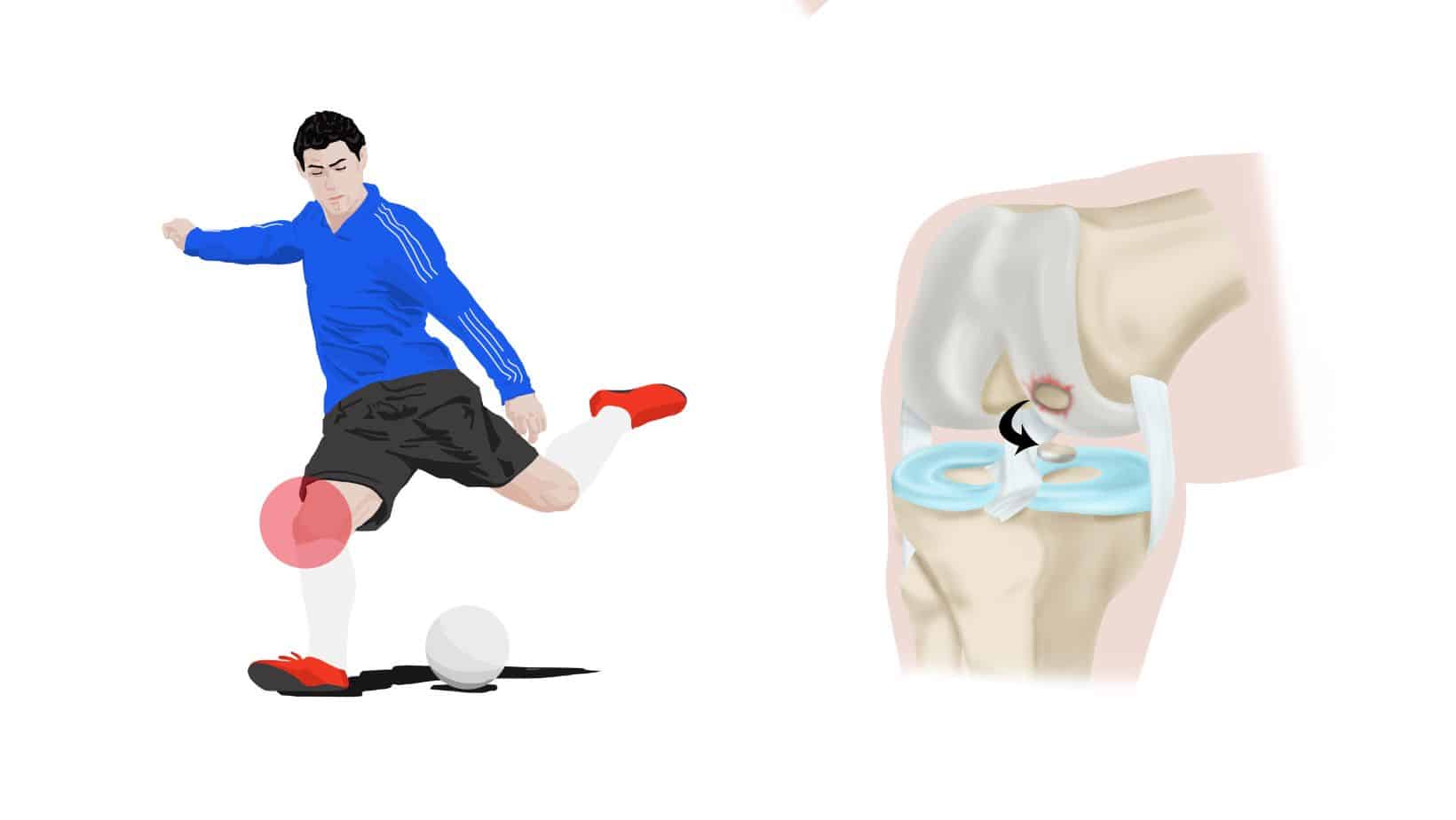
The knee is the most common joint affected by OCD, but it can affect any joint of the body. Other common sites include the elbow and ankles. Children and teenagers are more likely to have this condition and it is termed juvenile OCD, but it can still occur in adults.
When the blood flow is restricted the bone tissue will perish and can detach from the cartilage. This results in a fracture between the bone and/or cartilage in the joint, and fragmentation of bone and cartilage, this is called lesions. Therefore, loose bodies of bone and cartilage can be in the joint to cause symptoms.
It is pronounced os-tee-o-kon-dry-tis dis-uh-kanz, it is also known as OCD or JOCD for the juvenile form.
Pain will be felt in the affected joint and will be triggered by physical activity, this may be sports but can also be normal daily activities such as walking, descending stairs, or standing up from sitting. Often the affected joint will be swollen and may feel warmer than the other side. Loose bodies that have detached from the joint may block movement so the joint may not have its full range of mobility. In more advanced OCD people may also feel that the joint is unstable, or might “give way”, or that the joint catches.
Diagnosis should be by a physical therapist or specialist doctor and should include a thorough history of the presenting symptoms followed by a physical examination. This will be able to rule in or out many other causes of similar knee pain and symptoms. An orthopaedic test that is used for the diagnosis of osteochondritis dissecans is the Wilsons’s test. The final diagnosis is confirmed with radiology in the form of MRI or X-ray to show the separation of the cartilage and bone, and any loose bodies in the joint.
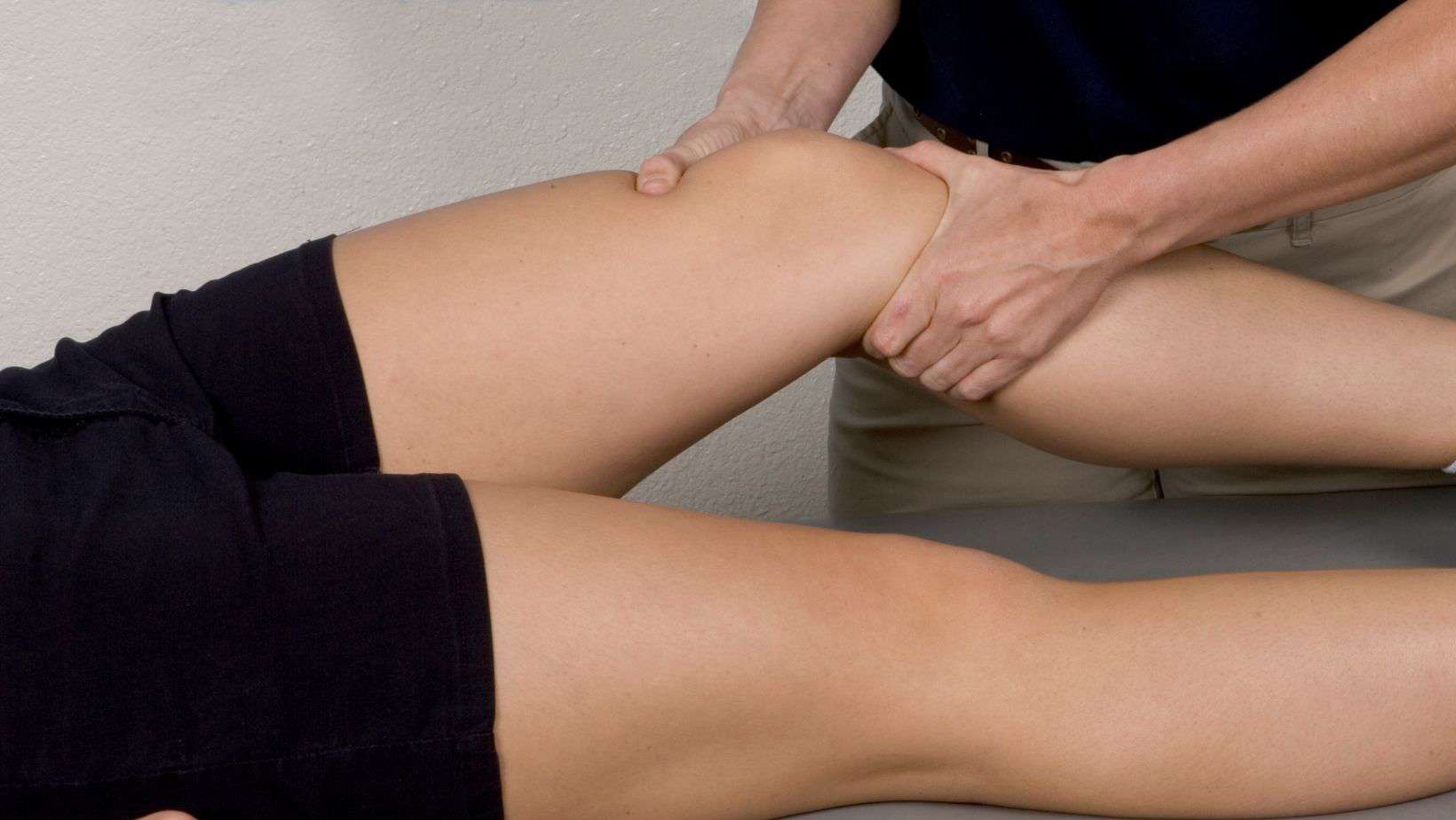
The knee is bent to 90º and the shin bone is rotated inwards, the knee is then straightened. A positive test is if pain is felt at about 70º of flexion on the inside of the knee.
There are 4 distinct stages of this pathology, categorised related to the International Cartilage Repair Society grading.
Is stable, there is the beginning of osteonecrosis in the bone, but the bone remains fully covered by intact cartilage.
Is stable, there is a further progression of the osteonecrosis, a osteonecrotic subchondral lesion develops and separation between the cartilage and bone begins.
Is unstable, an osteonecrotic fragment is formed but still attached to the cartilage and bone.
Is unstable, the osteonecrotic fragment becomes detached from the healthy bone.
Physiotherapy with James McCormack
This article is written by James McCormack, a Lower Limb Specialist who is an expert in treating Osteochondritis Dissecans.
This is not medical advice. We recommend a consultation with a medical professional such as James McCormack if you are experiencing any of the symptoms discussed in this article. James offers Online Physiotherapy Appointments weekly and face-to-face appointments in his London clinic.
Related Articles: Osteochondritis Dissecans Treatment