
Foot Bone or Joint Injury
Cuboid Syndrome
Read More >

Minute Read
|Posted 2 years ago
|Last updated: 28/08/2023
|by James McCormack

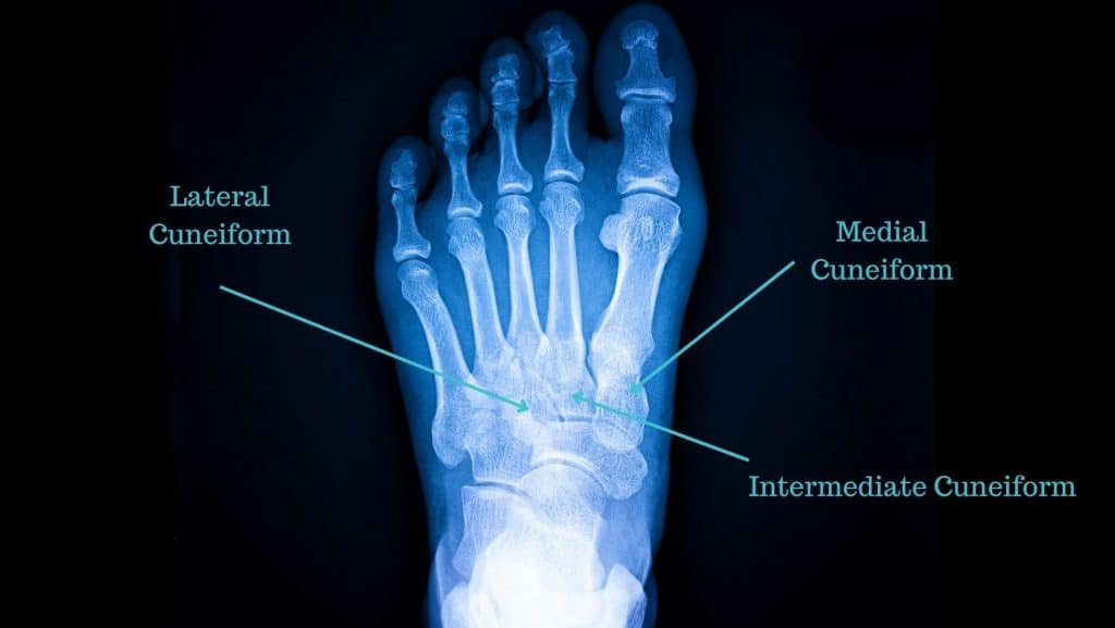
The cuneiform bones are a very small group of 3 bones. The lateral, middle and medial cuneiforms sit across the midfoot. The intermediate (middle) cuneiform is a wedge-shaped dome between the medial and lateral cuneiforms.
These are tightly packed and connected to the rest of the foot by some large ligaments, primarily to the 1st metatarsal and the Navicular Bone. As a result, injuries to the cuneiforms are rarely isolated but commonly a secondary consequence of another injury.
The Cuneiform bones provide attachment points for the Anterior Tibial Tendon, Peroneus Longus, Tibialis Posterior and Flexor Hallucis Longus.
It is more common to fracture or injure other structures in the midfoot than the cuneiform bones. A study on adult fractures concluded that cuneiform fractures comprise 0.1-0.5% of all adult fractures.
Other common injuries in the midfoot include:
The awareness of midfoot fractures has increased in recent times. This is due to a high-profile incident where American President Joe Biden had a cuneiform fracture. Here are the common types of Cuneiform fractures:
Cuneiform Avulsion Fracture occurs when the ligament or tendon detaches, pulling some bony fragments from the Cuneiform bone.
A non-displaced cuneiform fracture occurs when the bone remains intact and usually recovers with conservative treatment.
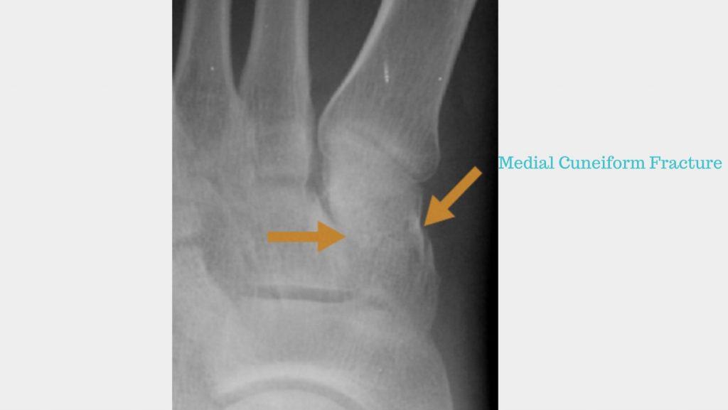
Middle Cuneiform Fracture – This type of hairline fracture of the cuneiform bone. It can be challenging to detect on an x-ray, and an MRI is often required.
Lateral cuneiform Fracture – This is an uncommon site of injury.
Cuneiform fractures are rare, and there is little evidence or case studies about them in the current literature. They are most often seen in high-speed incidents or as a result of overuse. Here are some causes from case studies.
In our experience, a cuneiform fracture takes 6-12 weeks to heal.
A cuneiform fracture is initially placed in a walker boot for 4-6 weeks. In the first instance, this is non-weight-bearing and progresses to weight-bearing.
Once the walker boot is removed, a period of Physical Therapy for 4-8 weeks commences. This protocol is not the case for all cuneiform fractures, and we advise seeing a Foot and Ankle Orthopedic Consultant for their opinion if you have a cuneiform fracture.
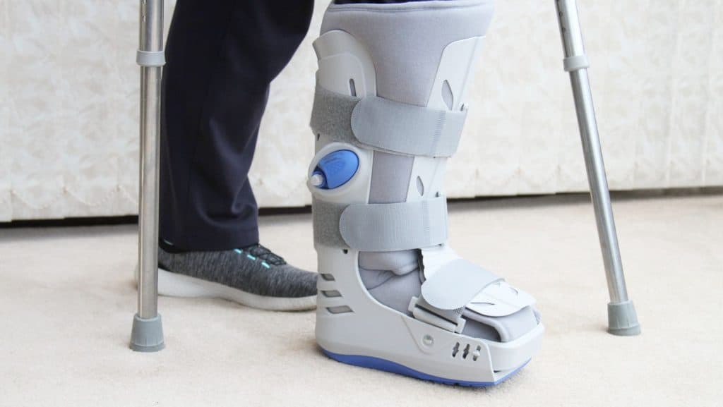
When wearing an air-cast walker boot, you can walk on a cuneiform fracture. It is not advised to walk barefoot with a cuneiform fracture.
Yes. Impact activities such as running can cause cuneiform fractures.
There are three cuneiform bones in the foot. These consist of the medial cuneiform, which is positioned on the foot’s inner side and is the largest of the three cuneiform bones. Next to the medial cuneiform is the intermediate cuneiform and the lateral cuneiform bone to the outside.
This is not medical advice. We recommend a consultation with a medical professional such as James McCormack. He offers Online Physiotherapy Appointments weekly.